Fluorescence Imaging
In vivo fluorescence imager
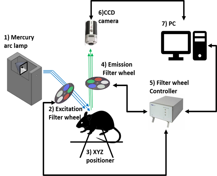
Schematic of the in vivo fluorescence imager
Portable in vivo fluorescence imager
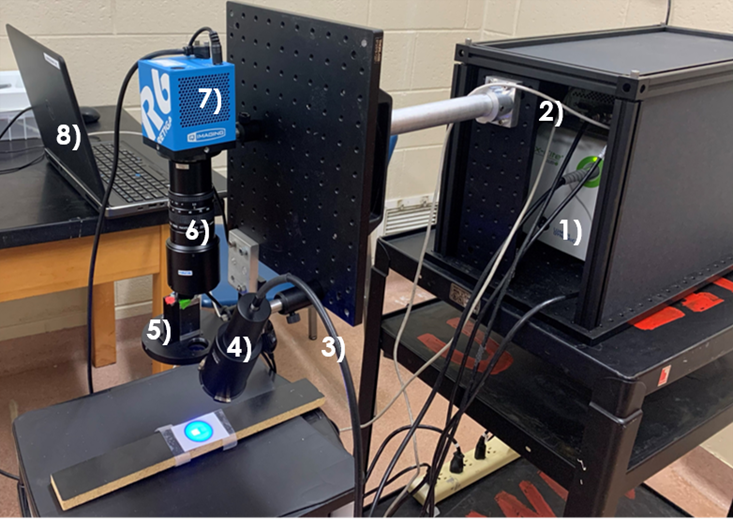
1. The excitation source: Nikon intensilight, 2. Filter wheel controller: 2-channle stepper motor, 3. Liquid light guide, 4. XYZ positioner, 5. Excitation filter wheel.
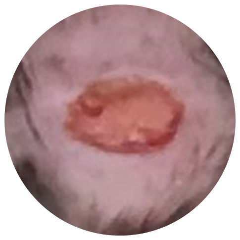
In vivo diabetic wound imaging
Diabetic wound
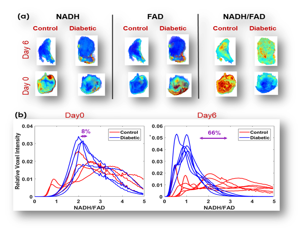
Top: Metabolic images of the wounds from a diabetic and control mouse.
Bottom: Redox state histogram of wounds in a diabetic vs. a nondiabetic changes over time.
Label-free Vessel Segmentation
Kidney
Raw 3D
3D Vasculature
Lung
Raw 3D
3D Vasculature
Airway
Hybrid
Irradiation injury
Terminal Points
Day 60
Day 90
Vessel Diameter
Day 60