Publications
Journal Publications
J44) M. Neghabi, P. Nategh, A. M. Stauffer, R. D. Blakely, and M. Ranji, "Elesclomol Diminishes Redox Imbalance in Peripheral Tissues of Mblac1 Knockout Mice," Journal of Biophotonics, vol. 19, no. 1, p. e70224, Jan. 2026, doi: 10.1002/jbio.70224.
J43) P. Nategh, M. Neghabi, J. F. Machi, I. Altilio, Y. Qi, A. A. Morales, D. H. Silvestre, D. R. Hernandez, N. Da Costa-Santos, A. G. Santana, M. Ranji, and C. O. Rodrigues, "Endothelial c-Myc and Doxorubicin-Induced Metabolic Alterations: A Multi-Organ Optical Imaging Study," Journal of Biophotonics, 2025, doi: 10.1002/jbio.70037.
J42) J. F. Machi, I. Altilio, Y. Qi, A. A. Morales, D. H. Silvestre, D. R. Hernandez, N. Da Costa-Santos, A. G. Santana, M. Neghabi, P. Nategh, T. L. Castro, J. P. Werneck-de-Castro, M. Ranji, F. S. Evangelista, R. I. Vazquez-Padron, E. Bernal-Mizrachi, and C. O. Rodrigues, "Endothelial c-Myc knockout disrupts metabolic homeostasis and triggers the development of obesity," Frontiers in Cell and Developmental Biology, vol. 12, July 2024, doi: 10.3389/fcell.2024.1407097.
J41) B. Ceyhan, P. Nategh, M. Neghabi, J. LaMar, S. Konjalwar, P. Rodriguez, M. K. Hahn, M. Gross, G. Grumbar, K. J. Salleng, R. D. Blakely, and M Ranji, "Optical Imaging Demonstrates Tissue-Specific Metabolic Perturbations in Mblac1 Knockout Mice" IEEE Journal of Translational Engineering in Health and Medicine, vol. 12, pp. 298-305, 2024, doi: 10.1109/JTEHM.2024.3355962, February 2024.
J40) S. Konjalwar, B. Ceyhan, O. Rivera, P. Nategh, M. Neghabi, M. Pavlovic, S. Allani, and M. Ranji, “Demonstrating drug treatment efficacies by monitoring superoxide dynamics in human lung cancer cells with time-lapse fluorescence microscopy,” J. Biophotonics, e202300331. DOI: 10.1002/jbio.202300331, October 2023.
J39) S. Mostaghimi, S. Mehrvar, F. Foomani, J. Narayanan, B. Fish, A.K.S. Camara, M. Medhora, and M. Ranji, “Vascular regression in the kidney: changes in 3D vessel structure with time post-irradiation,” Biomedical Optics Express, 13 (8), 4338-4352, DOI: 10.1364/BOE.464426, July 2022.
J38) F. Foomani, S. Mostaghimi, S. Mehrvar, S. Kumar, M. Ranji, “Optical Metabolic Imaging of Mitochondria Dysfunction in Heart and Kidney of Transgenic Mice”, IEEE JTEHM, DOI: 10.1109/JTEHM.2021.3104966, August 2021.
J37) S. Mehrvar, S. Mostaghimi, A. Camara, F. Foomani, J. Narayanan, B. Fish, M. Medhora, and M. Ranji, “3D Vascular Metabolic Imaging using Inverted Auto-fluorescence” J. Biomed. Opt. 26 (7), 076002, DOI: 10.1117/1.JBO.26.7.076002, July 2021.
J36) S. Gopalakrishnan, S. Mehrvar, S. Maleki, H. Schmitt, P. Summerfelt, A. M. Dubis, B. Abroe, T. B. Connor, J. Carroll, W. Huddleston, M. Ranji, and J. T. Eells, “Photobiomodulation Preserves Mitochondrial Redox State and is Retinoprotective in a Rodent Model of Retinitis Pigmentosa,” Scientific Reports, December 2020.
J35) S. Mehrvar, S. Mostaghimi, F. Foomani, J. Eells, B Abroe, and S. Gopalakrishnan, M. Ranji, “670nm Photobiomodulation improves the Mitochondrial Redox State of Diabetic Wounds” QIMS, doi: 10.21037/qims-20-522, July 2020.
J34) S. Mehrvar, F. Foomani, S. Shimada, C. Yang, N. Zheleznova, S. Mostaghimi, A Cowley, and M. Ranji, “The Early Effects of Uninephrectomy on Rat Kidney Metabolic State Using Optical Imaging,” Journal of Biophotonics, Doi: 10.1002/jbio.202000089, May 2020.
J33) S. Mehrvar, K.T. Rymut, F.H. Foomani, S.Mostaghimi, J.T. Eells, S. Gopalakrishnan, and M. Ranji, “Fluorescence Imaging of Mitochondrial Redox State to Assess Diabetic Wounds,” IEEE JTEHM, Doi: 10.1109/JTEHM.2019.2945323, October 2019.
J32) S. Mehrvar, M. Funding la Cour, M. Medhora, A. Camara, M. Ranji “Optical Metabolic Imaging for Assessment of Radiation-Induced Injury to Rat Kidney and Mitigation by Lisinopril,” Ann Biomed Eng, DOI: 10.1007/s10439-019-02255-8, April 2019.
J31) P. Kadamati, J.J. Sugar, B.J. Quirk, S. Mehrvar, G.G. Chelimsky, H.T. Whelan, T.C. Chelimsky, M. Ranji, “Near-infrared spectroscopy muscle oximetry of patients with postural orthostatic tachycardia syndrome,” Journal of Innovative Optical Health Sciences (JIOHS), Vol. 11, No. 6, DOI: 10.1142/S1793545818500268 , September 2018.
J30) Z. Ghanian, G.G. Konduri, S.H. Audi, A.K.S. Camara, M. Ranji, “Time‐lapse microscopy of oxidative stress demonstrates metabolic sensitivity of retinal pericytes under high glucose condition.,” Journal of Biophotonics, DOI: 10.1142/S1793545817500183, 2018, Aug. 2018.
J29) S. Lewis, T. Takimato, S. Mehrvar, H. Higuchi, A. Doebley, G. Stokes, N. Sheibani, S. IKeda, M. Ranji, and A. Ideka, “The effect of Tmem135 overexpression on the mouse heart,” PloS one, July 2018.
J28) P. Kadamati, J.J. Sugar, B.J. Quirk, S. Mehrvar, G.G. Chelimsky, H.T. Whelan, T.C. Chelimsky, M. Ranji, “Near-infrared spectroscopy muscle oximetry of patients with postural orthostatic tachycardia syndrome,” Journal of Innovative Optical Health Sciences (JIOHS), July 2018.
J27) Z. Ghanian, S. Mehrvar, N. Jamali, C.M. Sorenson, N.Sheibani, M. Ranji, “Time-lapse microscopy of oxidative stress demonstrates metabolic sensitivity of retinal pericytes under high glucose condition,” Journal of Biophotonics, doi: 10.1002/jbio.201700289, March 2018.
J26) M. Funding la Cour, S. Mehrvar, J. Heisner, M. Masoudi Motlagh, M. Medhora, M. Ranji, A. Camara, “Optical imaging of whole thorax irradiated rat hearts exposed to ischemia-reperfusion injury,” JBO, Vol. 23, No 1, DOI: 10.1117/1.JBO.23.1.016011, Jan 2018.
J25) M. Funding la Cour, S. Mehrvar, J. Kim, M. Zimmerman, J. Hong, M. Ranji, “Optical imaging for the assessment of hepatocyte metabolic state in ischemia and reperfusion injuries,” Journal of Biomedical Optics Express, Vol. 8, No. 10, September 2017.
J24) Z. Ghanian, G.G. Konduri, S.H. Audi, Amadou K.S. Camara and M. Ranji, “Quantitative optical measurement of mitochondrial superoxide dynamics in pulmonary artery endothelial cells,” Journal of Innovative Optical Health Sciences (JIOHS), Vol. 11, No. 1, June 2017.
J23) E. Aboualizadeh, M. Ranji, C.M. Sorenson, R. Sepehr, N. Sheibani, C. Hirschmugl, “Retinal oxidative stress at the onset of diabetes determined by synchrotron FTIR widefield imaging: Towards diabetes Pathogenesis,” Analyst, Feb. 2017.
J22) M. Ranji, M. MasoudiMotlagh, F. Salehpour, R. Sepehr, J.S. Heisner, R.K. Dash, A.K.S. Camara, “Optical Cryoimaging Reveals a Heterogeneous Distribution of Mitochondrial Redox State in ex vivo Guinea Pig Hearts and its Alteration during Ischemia and Reperfusion,” IEEE Journal of Translational Engineering in Health and Medicine (JTEHM), Vol. 4, July 2016. (Featured on IEEE EMBS website)
J21) Z. Ghanian, K. Staniszewski, N. Jamali, R. Sepehr, S. Wang, C.M. Sorenson, N. Sheibani, M. Ranji, “Quantitative Assessment of Retinopathy Using Multi parameter Image Analysis,” Journal of Medical Signals and Sensors, DOI: 10.1109/JTEHM.2016.2570219,Vol. 6, No.2, April 2016.
J20) S.H. Audi, A.V. Clough, S.T. Haworth, M. Medhora, M. Ranji, J.C. Densmore, E.R. Jacobs, “99mTc-Hexamethylpropyleneamine Oxime Imaging for Early Detection of Acute Lung Injury in Rats Exposed to Hyperoxia or Lipopolysaccharide Treatment,” Shock, DOI: 10.1097/SHK.0000000000000605, March 2016.
J19) A.W. Cowley, Jr., C. Yang, N. Zheleznova, A. Staruschenko, T. Kurth, K. Sadovnikov, A. Dayton, M. Hoffman, R. Ryan,M. Skelton, F. Salehpour, M. Ranji, and A. Geurts, “Evidence of the Importance of NOX4 in the Production of Hypertension in Dahl Salt-Sensitive Rats,” Hypertension, DOI:10.1161/HYPERTENSIONAHA.115.06.280, Dec. 2015.
J18) M. MasoudiMotlagh, M. Azimipour, J. Sugar, W.W Linz, G. Michalak, N. Seo, and M. Ranji, “Monitoring hemodynamic changes in stroke-affected muscles using near-infrared spectroscopy,” Journal of Rehabilitation and Assistive Technologies Engineering, Vol. 1, DOI: 10.1177/2055668315614195, Dec. 2015.
J17) F. Salehpour, Z. Ghanian, C. Yang, N.N. Zheleznova, T. Kurth, R. Dash, A.W. Jr Cowley, M. Ranji, “Effects of p67phox on the mitochondrial oxidative state in the kidney of Dahl salt-sensitive rats: Optical fluorescence 3D cryoimaging,” American Journal of Physiology Renal (AJP Rental), Vol. 309, No. 4, F377-F382, DOI: 10.1152/ajprenal.00098.2015, Aug. 2015.
J16) M. MasoudiMotlagh, R. Sepehr, N. Sheibani, C.M. Sorenson, and M. Ranji, “Optical cryoimaging of mitochondrial redox state in Bronchopulmonary-dysplasia injury models in mice lungs,” Quantitative Imaging in Medicine and Surgery, Vol. 5, No.1, Feb. 2015.
J15) Z. Ghanian, S. Maleki, F. Assadi-Porter and M. Ranji, “Optical imaging of mitochondrial redox state in rodent models with 3-iodothyronamine,” Journal of Experimental Medicine and Biology, Vol. 239, pp. 151- 158, DOI: 10.1002/jbio.201300033, Dec. 2013.
J14) Z. Ghanian, S. Maleki, F. Assadi-Porter and M. Ranji, ”Optical imaging of mitochondrial redox state in rodent models with 3-iodothyronamine,” Journal of Experimental Medicine and Biology, Vol. 239, pp. 151- 158, DOI 10.1002/jbio.201300033, Dec. 2013.
J13) R. Sepehr, S. Audi, K. Staniszewski, E. R. Jacobs, and M. Ranji, “Novel Flurometric Tool to Assess Mitochondrial Redox State of Isolated Perfused Rat Lungs after Exposure to Hyperoxia,” IEEE Journal of Translational Engineering in Health and Medicine, Vol. 1, No. 1, 1500210-1_1500210-10, Nov. 2013.
J12) Z. Ghanian, S. Maleki, C.M. Sorenson, SunYoung Park, N. Sheibani and M. Ranji, “Organ specific optical imaging of mitochondrial redox state in a rodent model of hereditary hemorrhagic telangiectasia-1,” Journal of Biophotonics, Vol. 6, No. 6, June 2013.
J11) R. Sepehr, S. Audi, S. Maleki, A.L. Eis, G.G. Konduri, M. Ranji, “Optical Imaging of Lipopolysaccharide-induced Oxidative Stress in Acute Lung Injury from Hyperoxia and Sepsis,” Journal of Innovative Optical Health Sciences (JIOHS), Vol. 6, No.3 June 2013.
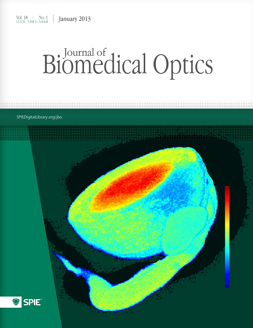
J10) S. Maleki, S. Gopalakrishnan, R. Sepehr, Z. Ghanian, H. Schmitt, J. Eells, M. Ranji, “Optical imaging of tissue mitochondrial redox state in rodent model of retinitis pigmentosa.” Journal of Biomedical Optics, Vol. 18, No. 1, Jan 2013. (Selected as cover page).
J9) K. Staniszewski, S. H. Audi, R. Sepehr, E. R. Jacobs, and M. Ranji, “Surface Fluorescence Studies of Tissue Mitochondrial Redox State in Isolated Perfused Rat Lungs.” Annals of Biomedical Engineering, Vol. 41, Jan. 2013.
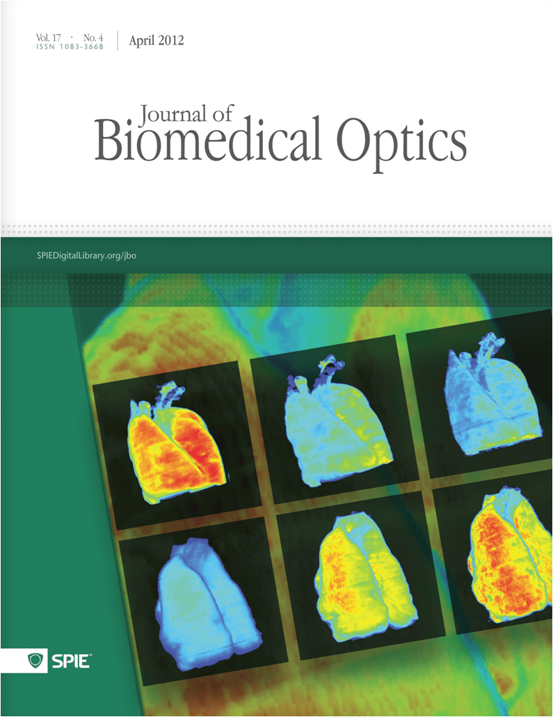
J8) R. Sepehr, K. Staniszewski, S. Maleki, E. R. Jacobs, S. Audi, M. Ranji, “Optical imaging of tissue mitochondrial redox state in intact rat lungs in two models of pulmonary oxidative stress.” Journal of Biomedical Optics, Vol. 17, No. 4, April 2012 (selected as cover page).
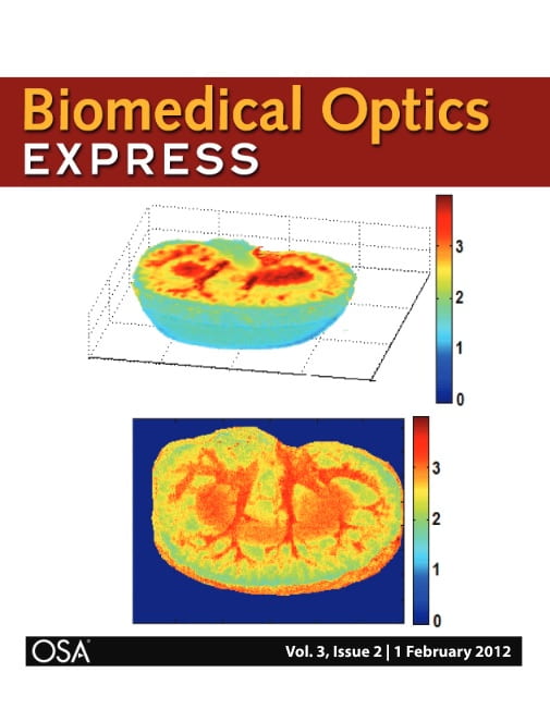
J7) S. Maleki, R. Sepehr, K. Staniszewski, N. Sheibani, C. M. Sorenson, and M. Ranji, “Mitochondrial redox studies of oxidative stress in kidneys from diabetic mice.” Journal of Biomedical Optics Express, Biomedical Optics Express, Vol. 3, No. 2, pp. 273-281, Feb. 2012 (Selected as cover page).
J6) M. Matsubara, M. Ranji, B. G. Leshnower, M. Noma, S. J Ratcliffe, and B. Chance, R. C. Gorman, J. H. Gorman III, “The Influence of Cyclosporine on Mitochondrial Function during Myocardial Ischemia and Reperfusion,” Annals of Thoracic Surgery, vol. 89, pp. 1532-7, May 2010.
J5) M. Ranji, S. Nioka, L. Z. Li, H. N. Xu, D. L. Jaggard and B. Chance, “Fluorescent Images of Mitochondrial Redox States of in Situ Mouse Hypoxic Ischemic Intestines,” Journal of Innovative Optical Health Sciences (JIOHS), Vol. 2, No. 4, pp. 365–374, Dec. 2009.
J4) L. Z. Li, H. N. Xu, M. Ranji, S. Nioka, and B. Chance, “Mitochondrial Redox Imaging for Cancer Diagnosis and Therapy,” Journal of Innovative Optical Health Sciences (JIOHS), Vol. 2, No. 4, pp. 325-341, Dec. 2009.
J3) M. Ranji, M. Matsubara, B. G. Leshnower, R. Hinmon, D. L. Jaggard, and B. Chance, R. C. Gorman, J. H. Gorman III, “Quantifying Acute Myocardial Injury Using Ratiometric Fluorometry,” IEEE Transaction on Biomedical Engineering (TBME), Vol. 56, No.6, pp. 1556-1563, May 2009.
J2) M. Ranji, S. Kanemoto, M. Matsubara, M. A. Grosso, J. H. Gorman III, R. C. Gorman, D. L. Jaggard, and B. Chance, “Fluorescence spectroscopy and imaging of myocardial apoptosis,” Journal of Biomedical Optics, Vol. 11, No. 6, Dec. 2006.
J1) M. Ranji, D. L. Jaggard, S. V. Apreleva, S. Vinogradov, and B. Chance, “Simultaneous fluorometery and phosphorometry of Langendorff perfused rat heart: ex vivo animal studies,” Optics Letters, Vol. 31, Issue 20, pp. 2995-2997, Oct. 2006.
Conference Proceedings
C69) P. Nategh, M. Neghabi, A. M. Stauffer, R. D. Blakely, and M. Ranji, "Elesclomol Diminishes Redox Deficit in Kidney of MBLAC1 Knockout Mice: A Fluorescence Imaging Study," in CLEO 2025, Technical Digest Series (Optica Publishing Group, 2025), paper JPS100_8.
C68) M. Neghabi, P. Nategh, B. Ceyhan, J. F. Machi, A. G. Santana, C. O. Rodrigues, and M. Ranji, "Investigating the Effects of Doxorubicin and Endothelial c-Myc Deficiency on Heart Metabolism using Fluorescence Cryo-Imaging," in 2024 46th Annual International Conference of the IEEE Engineering in Medicine and Biology Society (EMBC), Orlando, FL, USA, 2024, pp. 1-4, doi: 10.1109/EMBC53161.2024.11191576.
C67) P. Nategh, M. Neghabi, A. M. Stauffer, R. D. Blakely, and M. Ranji, "Elesclomol-Mediated Alterations of Liver Metabolism in the Context of Mouse Mblac1 Disruption," in 2024 46th Annual International Conference of the IEEE Engineering in Medicine and Biology Society (EMBC), Orlando, FL, USA, 2024, pp. 1-4, doi: 10.1109/EMBC53161.2024.11254869.
C66) M. Neghabi, P. Nategh, B. Ceyhan, S. Konjalwar, A. G. Santana, J. F. Machi, C. O. Rodrigues, and M. Ranji, "Quantifying the Effects of Chemotherapy-Induced Toxicity and Knockout of the Transcription Factor c-Myc on Mice Kidney Metabolism Using Optical Imaging," in CLEO 2024, Technical Digest Series (Optica Publishing Group, 2024), paper JTu2A.72.
C65) S. Konjalwar, B. Ceyhan, P. Nategh, M. Neghabi, S., Oscar Rivera, Shailaja Allani, M. Ranji, “Time-Lapse Fluorescence Microscopy to Study Superoxide Dynamics in Drug Treatment of Human Lung Cancer Cells,” Biomedical Engineering Society (BMES) Annual Meeting, Seattle, WA, Oct 2023.
C64) B. Ceyhan, J. LaMar, P. Nategh, M. Neghabi, S. Konjalwar, P. Rodrigues, M.K. Hahn, R. Blakely, M. Ranji, "Optical Imaging of Metabolic Dysfunction in Mice with a Genetic Deficiency in MBLAC1, a Risk Factor for Alzheimer’s Disease," IEEE EMBC conference, July 2023.
C63) P. Nategh, M. Neghabi, S. Mostaghimi, B. Ceyhan, S. Konjalwar, B. Fish, M. Medhora, and M. Ranji, “Using novel 3-D vascular metabolic imaging to assess effects of irradiation in lungs and kidney of rats”, LOPS Conference, June 2023.
C62) B. Ceyhan, S. Konjalwar, S. Mostaghimi, P. Nategh, M. Neghabi, B. Fish, M. Medhora, and M. Ranji, “Understanding why liver vasculature demonstrates higher resistance to radiation injury in female Wistar rat models,” LOPS Conference, June 2023.
C61) M. Ranji, "Optical imaging of tissue metabolism and vasculature,” LOPS Conference, June 2023.
C60) M. Dabagh, M. Ranji, "Prediction of ischemia reperfusion injury zone using in silico model of cardiac injury zone," the Conference on Lasers and Electro-Optics (CLEO), May 2023.
C59) S.Mostaghimi, S. Mehrvar, F. Foomani, J. Narayanan, A. Camara, B. Fish, M. Medhora, and M. Ranji, “Vascular Injury in Lung Vessels Post-radiation: A Dose and Time Response Case Study,” the Conference on Lasers and Electro-Optics (CLEO), May 2021.
C58) M. Ranji, F. Foomani, S. Mehrvar, S. Mostaghimi, N.Zheleznova, A. Cowley, “Optical imaging to assess the early metabolic response of rat kidney to uninephrectomy,” the Conference on Lasers and Electro-Optics (CLEO), May 2020.
C57) S. Mostaghimi, S. Mehrvar, F. Foomani, B. Abroe, J.T. Eells, M. Ranji, and S. Gopalakrishnan, “The Effect of NIR Light Treatment in Metabolic State of Diabetic Wounds,” BMES Annual Conference, Philadelphia, Oct. 2019.
C56) S. Mehrvar, F. Foomani, S. Mostaghimi, M. Medhora, A. Camara, and M. Ranji, "Radiation-induced Injuries to Rat Kidney Vasculature,” BMES Annual Conference, Philadelphia, Oct. 2019.
C55) F. Foomani, S. Mehrvar, S. Mostaghimi, S. Shimada, C. Yang, N. Zheleznova, Allen Cowley, and Mahsa Ranji, “The Early Effects of Uninephrectomy on Rat Kidney Metabolic State Using Optical Imaging,” BMES Annual Conference, Philadelphia, Oct. 2019.
C54) S. Mehrvar, K.T. Rymut, J.T. Eells, M. Ranji, and S. Gopalakrishnan, “Optical imaging for the metabolic redox state assessment of wound healing in diabetic mice,” Biophotonics Congress: Optics in the Life Sciences, Tucson, April 2019.
C53) S. Mehrvar, M. F. la Cour, M. Medhora, A. K. S. Camara, and M. Ranji, “Optical imaging for assessment of the impact of thoracic irradiation of hearts undergoing ischemia-reperfusion injury”, Engineering in Medicine and Biology Society (EMBC), 40th Annual International Conference of the IEEE, July 2018.
C52) S. Mehrvar, K. T. Rymut, J. T. Eells, M. Ranji, and S. Gopalakrishnan, “Optical imaging for the metabolic redox state assessment of wound healing in diabetic mice”, Symposium on Advanced Wound Care, June 2018.
C51) K. Sannagowdara, M. Malloy, W.L. Chen, B. Quirk, P.Kadamati, J. Sugar, M. Ranji, P. Monrad, J. Brown, and H. Whelan, “Cerebral oxygen saturation and cytochrome oxidase redox state in children with epilepsy: A pilot study - MULTICHANNEL NIRS for epilepsy seizure detection,” Clinical Neurophysiology, Volume 129, Pages e212-e212, May 2018.
C50) S. Mehrvar, K.T. Rymut, J.T. Eells, M. Ranji, and S. Gopalakrishnan, “When time does not heal wounds: optical imaging of diabetic wounds,” Three-Minute Thesis Competition (3MT), Milwaukee, April 2018.
C49) S. Mehrvar, M. Medhora, A.K.S. Camara, M. Ranji, “Optical cryoimaging for assessment of radiation-induced injury to rat kidney metabolic state,” SPIE BIOS, San Francisco, Feb. 2018.
C48) S. Mehrvar, M. la Cour, J. Kim, A. Martin, M. A. Zimmerman, J. Hong, and M. Ranji, “Optical imaging for liver transplant application,” 5th Annual Solid Organ Transplantation Research Symposium, Milwaukee, Fall 2017.
C47) Z. Ghanian, S. Mehrvar, N. Jamali, N. Sheibani, and M. Ranji, “A Comparison of retina endothelial cells and pericytes in metabolic sensitivity using time-lapse microscopy,” McPherson Eye Research Institute Poster Session, Madison, Fall 2017.
C46) S. Mehrvar, M.F. la Cour, M. Medhora, A.K.S. Camara, and M. Ranji, “Ischemia reperfusion in hearts: optical cryo-imaging,” Milwaukee Engineering Research Conference, Milwaukee April 2017.
C45) S. Mehrvar, M.F. la Cour, M. Medhora, A.K.S. Camara, and M. Ranji, “Optical Cryoimaging of Hearts during Ischemia and Reperfusion,” IEEE Larry Hause Student Poster Competition, Milwaukee March 2017.
C44) S. Mehrvar, Z. Ghanian, G. Kondouri, A.K.S. Camara, & M. Ranji, "Time-lapse microscopy of lung endothelial cells under hypoxia," In Proc. of SPIE, San Francisco, Feb. 2017.
C43) S. Bolin, G. Chen, M.M. Medhora, A.K.S. Camara, M. Ranji, “Optical imaging of mitochondrial redox state in irradiated vs. non-irradiated rat hearts during ischemia and reperfusion,” SPIE BIOS, San Francisco, Feb. 2016.
C42) A.W. Cowley Jr, C. Yang, N.N.Zheleznova, A.Staruschenko, T.Kurth, L. Rein, V. Kumar, K.Sadovnikov, A. Dayton, M. Hoffman, R.P. Ryan, M.M. Skelton, F.Salehpour, M. Ranji, A.Geurts, “Evidence of the importance of Nox4 in production of hypertension in Dahl salt-sensitive rats”, Hypertension, 2016.
C41) Z. Ghanian, G.K. Konduri, and M. Ranji, “Time lapse fluorescence microscopy of Reactive Oxygen Species (ROS) in vitro demonstrates a major role of complex IV in ROS generation,” RegionalBMES, Raleigh, Oct. 2015.
C40) M. Ranji, JJ. Sugar,E. Weiss, B. Quirk, H. Whelan, “Near Infrared spectroscopy (NIRS) of Cytochrome Oxidase,” SFN, Chicago, Oct. 2015.
C39) S. Bolin, G. Chen, M. Medhora, A.K.S. Camara, M. Ranji, “Optical imaging of mitochondrial redox state in irradiated vs. non-irradiated rat hearts during ischemia and reperfusion,” CTSI and Milwaukee Regional Research Forum, Oct. 2015.
C38) Z. Ghanian, A.G. Eis, G.K. Konduri, and M. Ranji, “Optical studies of oxidative stress in pulmonary artery endothelial cells,” SPIE BIOS, San Francisco, Feb. 2015.
C37) F. Salehpour, C. Yang, T. Kurth, A. W. Cowley Jr, andM. Ranji, “Optical cryoimaging of rat kidney and the protective effect of p67 in salt-induced hypertension”, SPIE BIOS, San Francisco, Feb. 2015.
C36) G. Konduri, M. Ranji,“Optical Studies of Oxidative Stress in Persistent Pulmonary Hypertension Cells,” Optical Molecular Probes,Imaging and Drug Delivery, 2015.
C35) A. W. Cowley Jr., F. Salehpour, C. Yang, T. Kurth, M. Ranji, “Cryofluorescence 3D imaging shows mutation p67phox improves metabolic function and reduces oxidative stress in the renal medulla of Dahl salt-sensitive rats”, American Heart Association Conference, High Blood Pressure Research, San Francisco, CA, Sept. 2014.
C34) T. R. Schaid A. H. Abdelhafeez, M. Ranji, R. Love, S. Audi, S. Kaul, F. Bashiri, E. MasoudiMotlagh, F. Salehpoor, E. Jacobs, J. C. Densmore, “Surface Fluorescence Studies Of Tissue Mitochondrial Redox State In Ex-vivo Lung Perfusion,” Shock 37th annual Conference, June 2014.
C33) Z. Ghanian, K. Staniszewski, R. Sepehr, C.M. Sorenson, N. Sheibani, and M. Ranji, “Cytometric classification of retinopathic injury,” SPIE Proceeding of SPIE BIOS, San Francisco, Feb. 2014.
C32) W.W. Linz, G. Michalak, M.M. Motlagh, N. Seo, M. Ranji, “Blood volume and oxygenation changes in the skeletal muscles of stroke patients measured using non-invasive surface near-infrared spectroscopy,” SPIE Proceeding of SPIE BIOS, San Francisco, Feb. 2014.
C31) M. Ranji, Z. Ghanian, F. Atry, S. Frye, R. Pashaie, S. Audi, “Optical Instrumentation and the Image Cytometry of Lung and Eye Injuries: Studies in the Rodent Model,” OSA Biomedical Optics, 2014.
C30) A.W. Cowley, F Salehpour, C Yang, T Kurth, M Ranji, “Cryofluorescence 3D Imaging Shows Mutation of p67phox Improves Metabolic Function and Reduces Oxidative Stress in the Renal Medulla of the Dahl Salt-sensitive Rat,” Hypertension 64 (suppl_1), A665-A665, 2014.
C29) J.T. Eells, S. Gopalakrishnan, H. Schmitt, S. Maleki, A. Dubis, J. Carroll, M.Ranji, “Photobiomodulation Protects Retinal Mitochondria and Retinal Function in a Rodent Model of Retinitis Pigmentosa,” ARVO Asia 2013.
C28) Z. Ghanian, Mohammad MasoudiMotlagh, S. Maleki, Z. Bolandnazar, F. Assadi-Porter, M. Ranji, “Optical Redox Imaging of Metabolic Dysfunction in Polycystic Ovary Syndrome,” BMES Annual Conference, Seattle, Sept. 2013.
C27) W. Linz, G. Michalak, M. MasoudiMotlagh, N. Seo, M. Ranji, “Hand function assessment of stroke patients using non-invasive surface near-infrared spectroscopy,” Great Lake Biomedical Conference, GE Healthcare, Milwaukee, April 2013 (awarded third place).
C26) M. MasoudiMotlagh, S. Maleki, F. Assadi-Porter, M. Ranji, “Optical Redox Imaging to Monitor Metabolic Dysfunction in Polycystic Ovary Syndrome,” 2013 Great Lake Biomedical Conference, GE Healthcare, Milwaukee, April 2013.
C25) Z.Ghanian, S.Maleki, C.M. Sorenson, N.Sheibani, M. Ranji, "Optical Imaging of Cellular Redox State Related to Retinopathy Dysfunction in Endoglin Heterozygous (Eng+/-) Mice," UWM CEAS Research Day, April 2013.
C24) Z. Ghanian, S. Maleki, S. Gopalakrishnan, R. Sepehr, J. T. Eells and M. Ranji, “Optical Imaging of Oxidative Stress in Rodent Model of Retinitis Pigmentosa,” Proceeding of SPIE BIOS, San Francisco, Jan. 2013.
C23) R. Sepehr, K. Staniszewski, E. R. Jacobs, S. Audi, and M. Ranji, “Fluorometry of ischemia reperfused rat lungs in vivo,” Proceeding of SPIE BIOS, San Francisco, Jan. 2013.
C22) JT Eells, S Gopalakrishnan, S Maleki, M Ranji, B Abroe, H Schmitt, P Summerfelt, A Dubis, J Carroll, “Photobiomodulation preserves mitochondrial redox state in a rodent model of retinitis pigmentosa,” Mitochondrion, 2013.
C21) R. Sepehr, S. Maleki, A.L. Eis, G.G. Konduri, M. Ranji, “Optical Imaging of Hyperoxic Lung Injury,” Photonics Global Conference (PGC), Singapore, Dec. 2012.
C20) Z. Ghanian, S. Maleki, S. Gopalakrishnan, C.R. Sorenson, N. Sheibani and M. Ranji, “Optical imaging of Oxidative Stress in Diabetic Retinopathy,” BMES Annual Conference, Atlanta, Oct. 2012.
C19) M. Ranji, "Quantitative Deconvolution and Tracking of Differentiating Stem Cells" TERMIS, Vienna, September 2012.
C18) K.Staniszewski, R. Sepehr, S. Maleki, C.M. Sorenson,N. Sheibani, and M. Ranji “Automated Evaluation of Retinopathies Using Image Cytometry,” Proceedings of Data Analysis and Modeling Retina in Health and Disease, Lisbon, Feb. 2012.
C17) R. Sepehr, K. Staniszewski, E. R. Jacobs, S. Audi, and M. Ranji, “Optical studies of tissue mitochondrial redox in isolated perfused rat lungs,” Proceedings of SPIE 8207D, San Francisco, Jan. 2012.
C16) S. Maleki, R. Sepehr, K. Staniszewski, N. Sheibani, C.M. Sorenson and M. Ranji, “Optical cryoimaging of kidney mitochondrial redox state in diabetic mice models,” Proceedings of SPIE 8225, San Francisco, Jan. 2012.
C15) K. Staniszewski, R. Sepehr, C. M. Sorenson, N. Sheibani,and M. Ranji, “Classification of retinopathic injury using image cytometry and vasculature complexity,” Proceedings of SPIE, San Francisco, Jan. 2012.
C14) R. Sepehr, K. Staniszewski, M. Ranji, and S. Maleki, “Optical Cryoimaging Of Kidney Mitochondrial Redox State And The Effect Of Bcl-2 Family Expression,” Proceeding of BMES Annual Meeting, Hartford, Oct. 2011.
C13) R. Sepehr, S. Audi, K. Staniszewski, S Maleki, and M. Ranji, “Fluorescence Spectroscopy and Cryoimaging of Rat Lung Tissue Mitochondrial Redox State,” Proceedings of SPIE 80870, Munich, doi:10.1117/12.890019, June 2011.
C12) M. Ranji, D. Calzolari, R. Augustin, and J.H. Price, “Is Image Cytometry Possible with Deconvolved Fluorescence Images?,” OSA, 2010.
C11) M. Ranji, D.Calzolari, R. Agustin, and J.H. Price, “Quantitative analysis of 3D Fluorescence Images,” Cytometry Development Workshop (CDW), Asilomar, Oct. 2009.
C10) M. Ranji, D.Calzolari, N. Prigozhina, K.A. Wei, M. Mercola, J.H. Price, “Automated Tracking of Migration and Differentiation of Fluorescently Labeled Human Embryonic Stem Cells,” Proceedings of SPIE, Bios, Jan. 2009.
C9) M.Ranji, D. Calzolari, and J.H. Price, “Quantitative analysis of deconvolution methods for fluorescence microscopy images,” Invited, Asilomare conference SSC, Oct. 2008.
C8) M.Ranji, M. Matsubara, B.G. Leshnower, R.Hinmon, D.L. Jaggard and B.Chance, R.C. Gorman and J. H. Gorman III, “Optical Biopsy of Apoptosis in Ischemic Myocardium with Fluorometry,” Proceedings of AHA (American Heart Association), Nov. 2007.
C7) M. Ranji, M. Matsubara, M. Grosso, D.L.Jaggard and B.Chance, R.C. Gorman and J. H. Gorman III, “Fluorescence Spectroscopy to Assess Apoptosis in Myocardium,” Proceedings of SPIE, Vol. 6438, pp. 64380J1- 64380J4, Feb. 2007.
C6) M. Ranji, M. Matsubara, M.A. Grosso, D.L Jaggard, B. Chance, R.C. Gorman, J.H. Gorman III, " Fluorescence spectroscopy to assess apoptosis in myocardium,” Proceedings of SPIE 64380J, San Jose, Jan. 2007.
C5) M. Ranji, D.L.Jaggard and B. Chance, “Fluorescence Spectroscopy of Perfused Rat Heart: a Fluorometer Study,” Proceedings of the IEEE Bioengineering Conference, pp. 203-204, April 2006.
C4) M. Ranji, L.Z. Li, J. Glickson, and B. Chance “Optical cryoimaging of tumor metabolism and aggressiveness,”The Eunice and Irving Leopold Annual Scientific Symposium, March 2006.
C3) M. Ranji, B. Chance, L. Moon, J. Gorman, L. Li, and C. Thompson, “Apoptosis Gives an Intrinsic Optical Signal,” OSA Biomedical Optics Proceedings, March 2006.
C2) L. Zhou, M. Ranji, and B. Chance, “Novel Fluorochromes for Functional Imaging of Cancer,” OSA Biomedical Optics proceedings, March 2006.
C1) M. Ranji, D.L.Jaggard and B.Chance, “Observation of mitochondrial morphology and biochemistry changes undergoing apoptosis by angularly resolved light scattering and cryoimaging,” Proceedings of SPIE, Vol. 6087, pp. 60870K1 - 60870K9, Jan. 2006.