1/11/2024
Art of Science: Brain Cell Galaxy
Award-winning Photograph Showcases Neuroscience
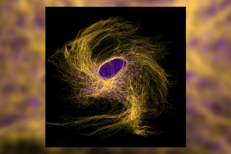
To learn more about Alzheimer’s disease, Peter Rodriguez, a doctoral student in the Charles E. Schmidt College of Science, takes microscopic images of cells in the brain of genetically altered worms and mice to study a previously unstudied gene. One of those images, called Brain Cell Galaxy, recently earned second place in the Division of Research Art of Science photography contest.
“It’s truly an honor to have research that we spend so much time on gain more attention through this Art of Science contest, as this not only brings attention to very important research, but also to the work we're doing in our lab specifically,” Rodriguez said. “I think that by combining art and science, it helps get the word out.”
The winning image shows a brain cell type called an astrocyte, said Rodriguez, which was cultured in a dish from brain tissue of a mouse. Astrocytes, named for their celestial appearance, are critical for brain health and help prevent the breakdown of tissue in the brain, such as in the case of Alzheimer’s, he said.
For his research, Rodriguez studies how the gene swip-10, also known as Metallo-Beta-Lactamase Domain Containing 1 (MBLAC1) in humans, plays a role in brain disease. He’s working with his advisor Randy Blakely, Ph.D., executive director of the Stiles-Nicholson Brain Institute, the David J.S. Nicholson Distinguished Professor in Neuroscience, and a professor of biomedical science in the Charles E. Schmidt College of Medicine, who discovered the swip-10 in 2012. Mblac1 has recently been implicated as a risk factor for Alzheimer’s “To get a better understanding of how this pathway is working, multiple approaches such as the use of genetic model animals have been critical. ” Rodriguez said.
One way to do that, he said, is to take cells from the mouse brain tissue, fix the tissue to a piece of glass, then stain it with fluorescent antibodies and examine it under a microscope. By staining the slide, the researchers can visualize different parts of the cell, their shape, size — called morphology — and health as well as the protein responsible for making the gene of interest. In his winning image, stained yellow is the inner, supportive skeleton of the cell and the nucleus, where DNA is located, is stained purple, Rodriguez said.
“Using these types of imaging approaches, we can study how genetics influence the morphology of important cell types, and how their shape might link to their function, giving us clues to brain health,” he said.
Rodriguez, a Florida native, received his bachelor’s degree in biology from Barry University, Miami, in 2016. After graduating, he was a research technician with Blakely studying nematodes, a type of microscopic worm used in Blakely’s lab to study brain diseases. During this time, he said he discovered his true love of neuroscience, which is why decided to start the doctoral program at Florida Atlantic. Studying the complexity of the brain feels like a quest, he said. “I can study this thing that makes us individuals, and then also, of course, hopefully, we can ultimately help people with our research,” he said.
For Rodriguez, sharing the beauty of science is important to help inspire young people, he said. “I never saw these types of images when I was younger and maybe it could inspire someone coming up, not knowing where they want to go in life. For an undergraduate student or high schooler, it could be really impactful.”
Art of Science
The 7th annual Art of Science exhibition was a huge success. If you missed it, here's a recap as well as a virtual walkthrough.
Recap
Walkthrough
Be a part of next year's fun and submit now to the 8th annual Art of Science contest.
View all Art of Science winners here.
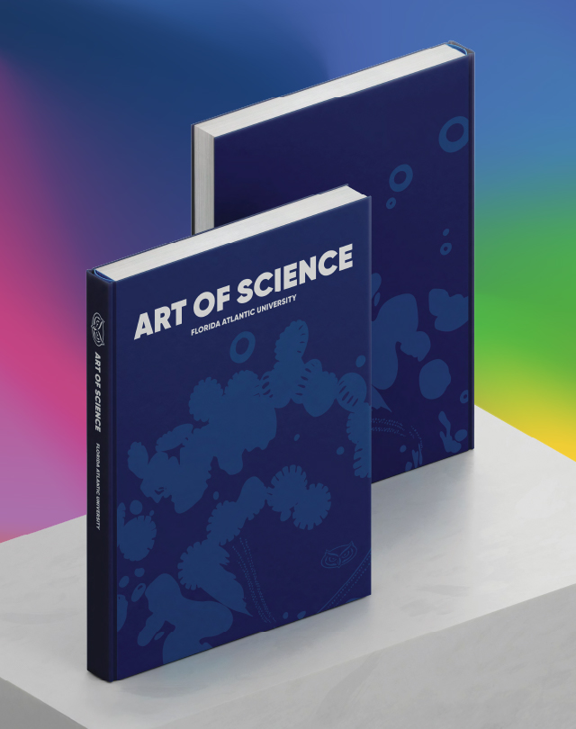
Get your copy of the commemorative Art of Science coffee-table book ...
All of the research
All of the pictures
All of the stories
All yours.