TIE: Student in the Lab: Thresher Vertebra
Photo by Jamie Knaub, graduate student,
Charles E. Schmidt College of Science
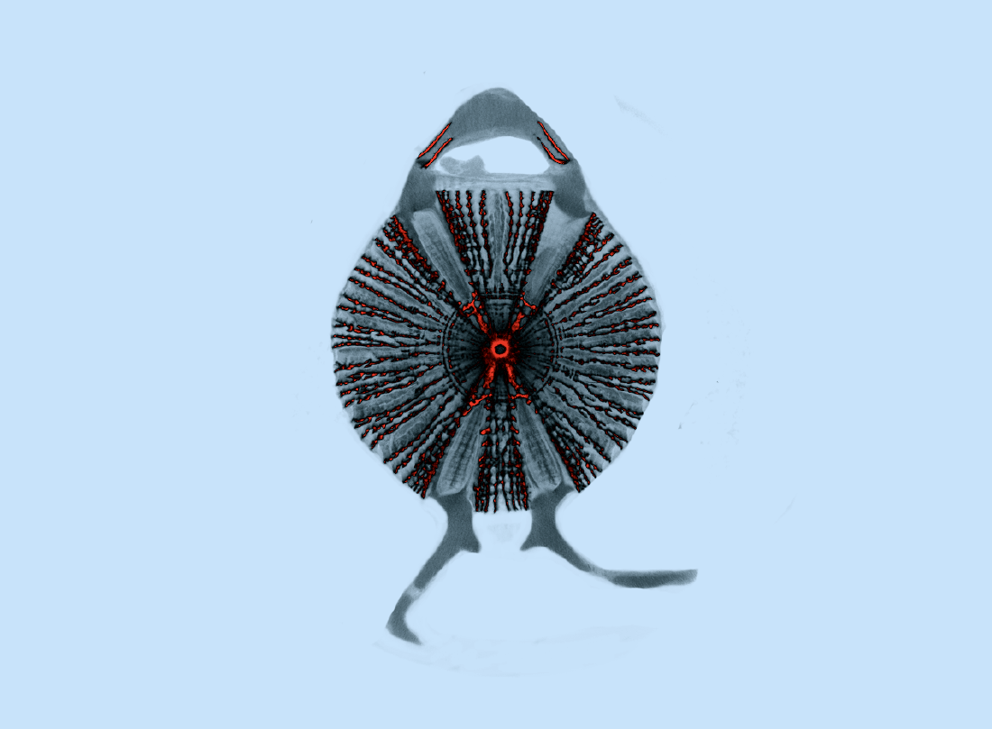
Thresher vertebra: Micro-CT image of a cross section of a thresher shark (Alopias vulpinus) vertebra. Sharks have mineralized cylindrical vertebrae with a calcified internal architecture. We can use micro-CT (computed tomography) imaging which relies on X-rays to visualize the internal mineral structures. The radiating lines from the center are called lamellae which sometimes branch at intersections termed nodes. Thresher sharks have the highest counts of lamellae and nodes which are believed to contribute to the stiffness and toughness of the vertebral cartilage. Here, the most mineralized portions are colored red. Additionally, the projections at the top and the bottom of the vertebra are the neural arch and hemal arches respectively. Image taken using Bruker Skyscan 1173 micro-CT scanner at FAU High School Owls Imaging Lab.