Honorable Mention: Sharktography
Photography by Dawn Raja Somu, doctoral student, candidate or graduate student,
Charles E. Schmidt College of Science
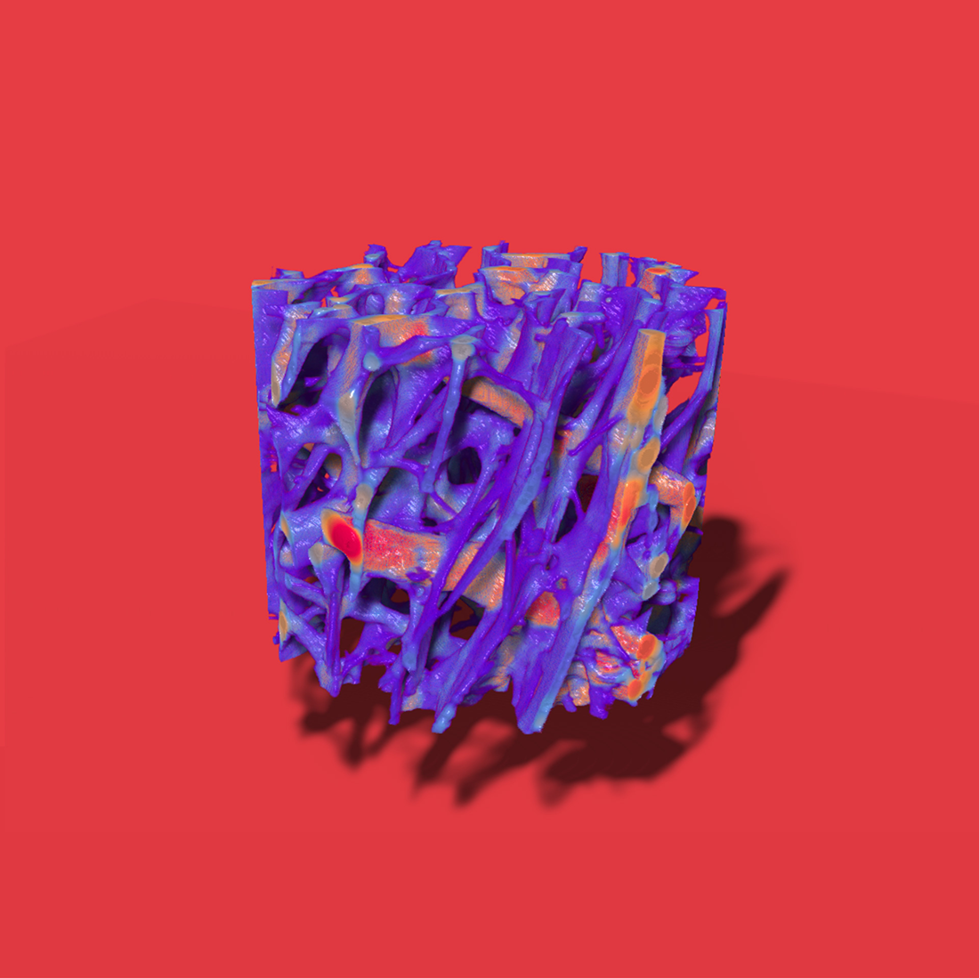
Mapping of the contours of shark vertebral cartilage: The endoskeleton of sharks is made up of cartilage, rather than bone. Apatite mineral, collagen fibers, and sugars serve as a framework for the strong and flexible material that makes up shark cartilage. The arrangement of these components in the fibers of shark vertebral cartilage affect how it responds to mechanical stress. We examine and characterize the micro- and nanoscale properties of shark cartilage to figure out how its structure influences macroscale function and performance. Presented here is a 3D model of mineralized vertebral cartilage of a blacktip shark (Carcarhinus limbatus) in the nanoscale using synchrotron X-ray nanotomography (SR-nanoCT – like a CT scan, but with stronger X-rays and better resolution). Here, the thickness of the cartilage is correlated with the colors on the 3D model. The thicker red regions are striations that run in the opposite direction of most fibers and are hypothesized to serve as struts that support the complex bending movements that sharks undergo while swimming. Insight into ultrastructure of a natural composite material such as shark cartilage can help in the design of bioinspired materials with specialized mechanical properties for applications in areas such as tissue engineering and materials for marine propulsive robots.
Mentor: Vivian Merk, Ph.D., assistant professor, Charles E. Schmidt College of Science