Mouse Model
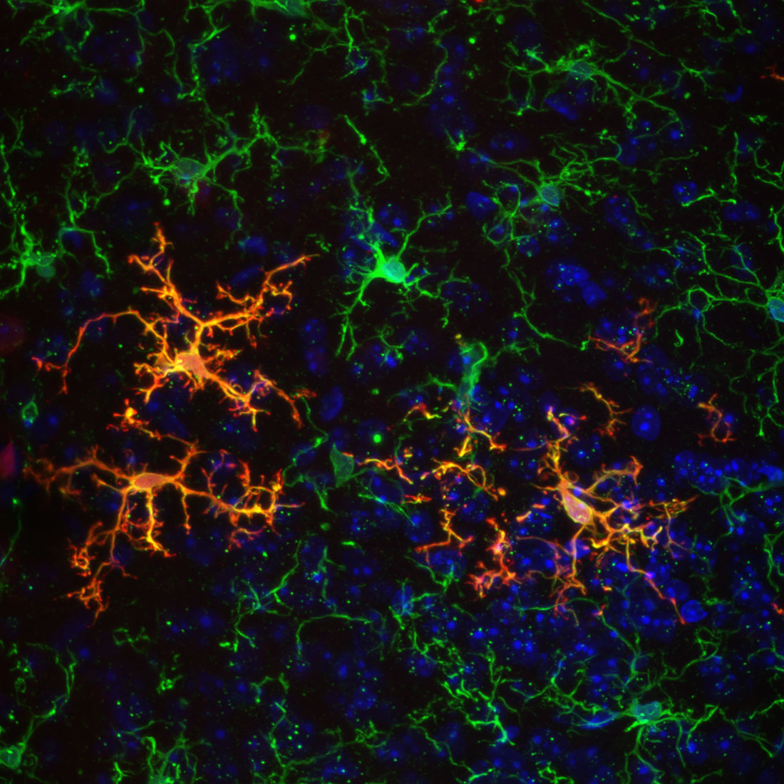
Interleukin-1 is an inflammatory molecule, called a cytokine, which is highly important for all infection, neurodegenerative disease, and its elevated levels are associated with cognitive decline and mood disorders. Understanding where and what cell expresses IL-1 in the brain allows us to detail where it is influencing neural circuits. FAU researchers generated a novel mouse model where all IL-1 expression is tracked by a red fluorescent molecule and we gain genetic access to each of these cells. In this image, cell nuclei (blue), the immune cell of the central nervous system called microglia (green), and IL-1 expressing cells (red) are shown. Notice how the red and green are overlapping making a yellow color. This means that IL-1 is primarily expressed by microglia. By tracking where IL-1 is expressed in the brain using this method, we can see which cells emit this immune signal during any adverse challenge to the brain such as infection, disease, and stress to understand which neural circuits and behaviors can be influenced by IL-1.