Pathology Progression
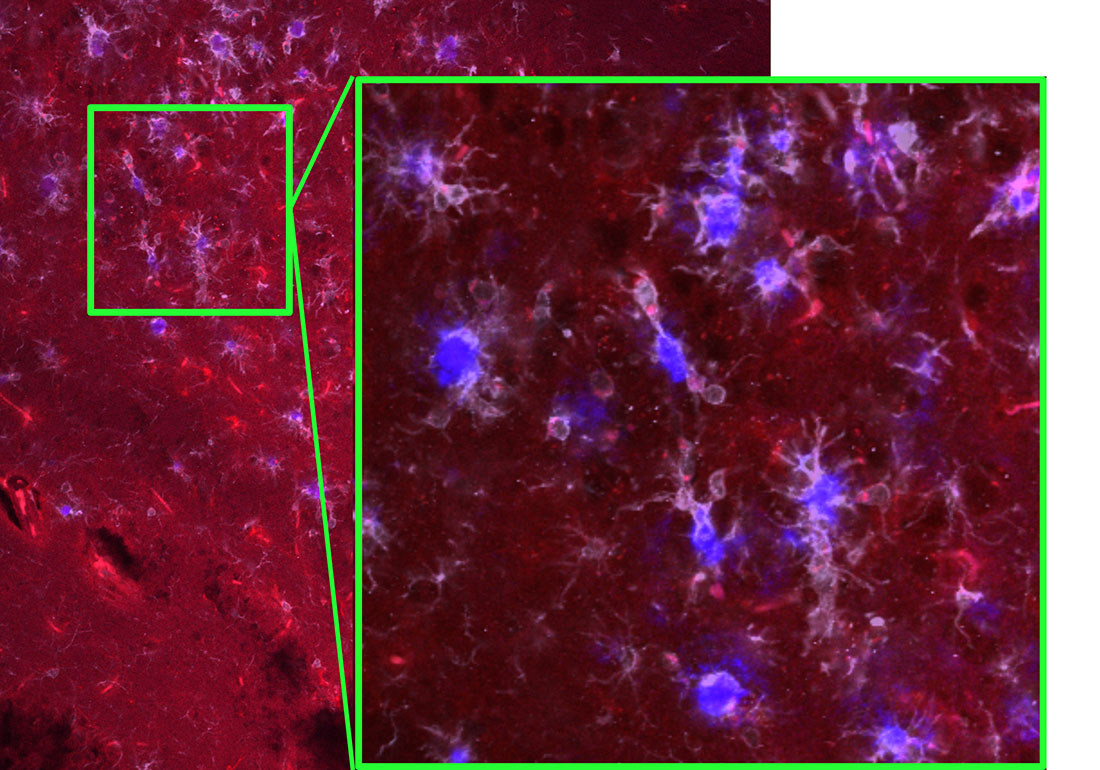
Aged 5xFAD Alzheimer's Mouse Hippocampus - Hippocampal brain sections of a transgenic Alzheimer’s mouse model of a 6-month-old mouse were investigated through IHC antibody staining such that the final antibody added will reflect a colored wavelength: Amyloid Beta Plaques (blue: Amylo Glo), IL1R1 (red: tdTom), Microglia (far red/gray: IBA1). At this stage in the mouse's lifespan Alzheimer's Disease pathology has progressed to the point of causing behavioral changes and cognitive deficits. Microglia, the brain's resident immune cell, form structures around the plaques with their cell processes as amyloid beta is accumulated during Alzheimer's pathology progression. The zoomed-in box shows a line of microglia (in gray); this formation is referred to as rod-like microglia and is a previously reported response to neuron cell death. Images were captured on a confocal microscope by creating a z-stack image (multiple layers pressed together) and edited on Biorender.