1/29/2025
Art of Science Announces Third-Place Winner
FAU Doctoral Student’s Eye-Catching Submission Captures Inner Beauty of the Brain
Great advancements in microscope technology enable leaps in neuroscience discovery – and they give us an inside view of the remarkable beauty of the brain.
Marianne Monet, a doctoral student at Florida Atlantic’s Stiles-Nicholson Brain Institute (SNBI) under the mentorship of Ning Quan, Ph.D., studies how the immune system affects the brain during childhood, and how these effects can influence behavior and choices later in life.
“In our research, we’ve discovered that multiple brain regions express the neuronal IL-1R1 protein,” Monet said. “We also identified that neuronal IL-1R1 plays a crucial role in social interaction deficits and stress exposure in early life.”
If a child faces frequent or long-term immune challenges, it activates the immune system, which can disrupt normal brain development. This disruption may affect brain areas essential for social behaviors, like forming and maintaining friendships, potentially making these social interactions more challenging as they grow older, Monet said.
It was during an early phase of this project that Monet captured the image that earned her third place in this year’s Art of Science contest.
“The first step of my project was to identify which brain regions and cell types express neuronal IL-1R1,” she said. “To do this, I mapped IL-1R1 expression across different brain areas in developing mice. During this initial mapping, I came across an eye-catching structure expressing IL-1R1 and captured the image.”
“Eye-catching” is the operative phrase. Using an epi-fluorescence microscope, the blood vessels and neurons that contain 1L-1R1 appear as a red, oblong shape surrounded by green-pigmented neurons that lack the target protein. The result looks uncannily like an eye staring back at the viewer from the inner reaches of a young mouse cerebellum.
“As a neuroscientist, I’m constantly fascinated by images of the brain,” Monet said. “This image showcases the beauty of the brain. It’s a remarkable organ capable of performing millions of functions simultaneously in such a short period of time.”
The epi-fluorescence microscope was essential to Monet’s investigation because it allowed her to quickly focus and scan brain regions, saving valuable time during imaging sessions. Its high-resolution magnification can capture both broad and detailed views of brain structures – as exemplified by her award-winning submission. And it can precisely focus on different depths within a tissue, “which is important for imaging complex, three-dimensional structures like the brain,” she said.
As her project progressed, Monet’s focus shifted to the hippocampus, so she didn’t have the opportunity to investigate the unique cerebellar structure featured in her image.
“I thought it was worth sharing, hoping it would spark curiosity and excitement in others about the wonders of the brain,” she said.
Note: Monet is a member of the elite International Max Planck Research School for Synapses and Circuits, a partnership between Florida Atlantic and Max Planck Florida Institute for Neuroscience, which is located adjacent to SNBI on FAU’s Jupiter Campus.
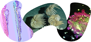
Art of Science
Entries are open for the 8th annual Art of Science contest.
View the 7th annual Art of Science Virtual Tour
View all Art of Science winners here.
Get your copy of the commemorative Art of Science coffee-table book ...
All of the research
All of the pictures
All of the stories
All yours.