Honorable Mention: Seagrass Root
Charles E. Schmidt College of Science
Mentor: Marguerite Koch-Rose, Ph.D.
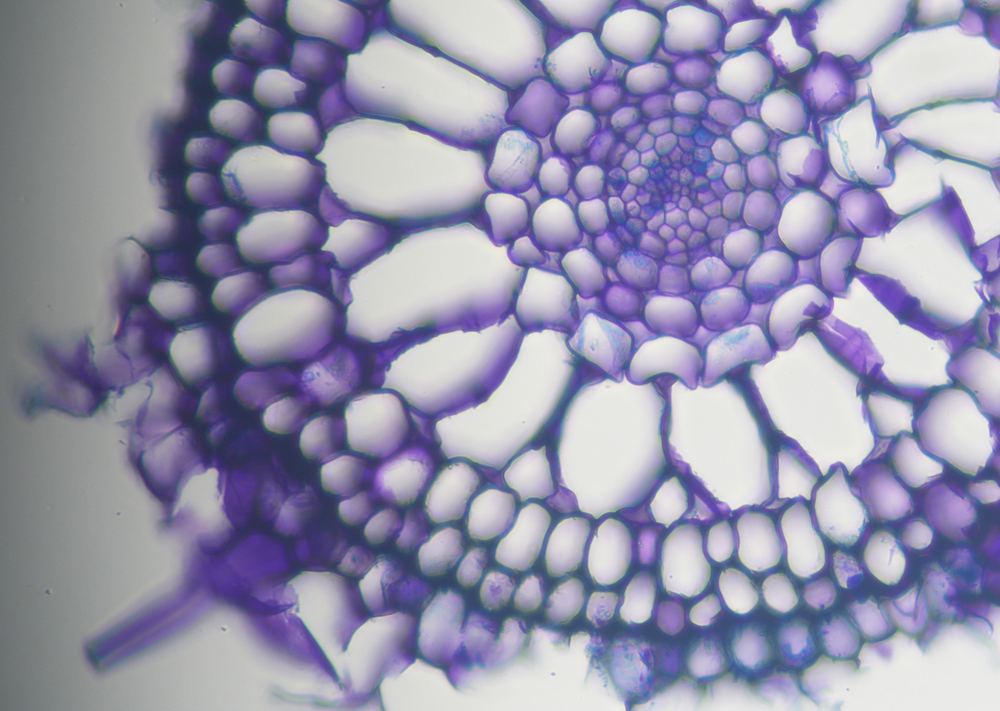
This transverse cross section reveals the elegant internal anatomy of a turtle grass (Thalassia testudinum) root, an unseen world beneath Florida Bay’s iconic meadows. Prominently featured in this microscopy image are large aerenchyma, or airspace tissues, that form a vital internal oxygen “highway,” transporting photosynthetically derived oxygen from the leaves to the roots. In the sediments where seagrasses grow, oxygen is a scarce but essential resource. These airspace structures are key to avoiding stress associated with low oxygen and enabling respiration where roots cannot rely on the surrounding environment. The root is stained with Toluidine blue, allowing us to determine what compounds compose seagrass cell walls. Cross sections like this offer insight into how anatomy supports function. Seagrasses are vital ecosystem engineers, stabilizing sediments, supporting biodiversity and sequestering carbon. By unraveling the physiological strategies used to survive in challenging environments, it deepens an understanding of resilience and informs efforts to protect and restore these coastal habitats in the face of accelerating change and human impact. This image was taken at the Berlin Family Bioimaging Lab at FAU Lab Schools Marcus Research and Innovation Center.