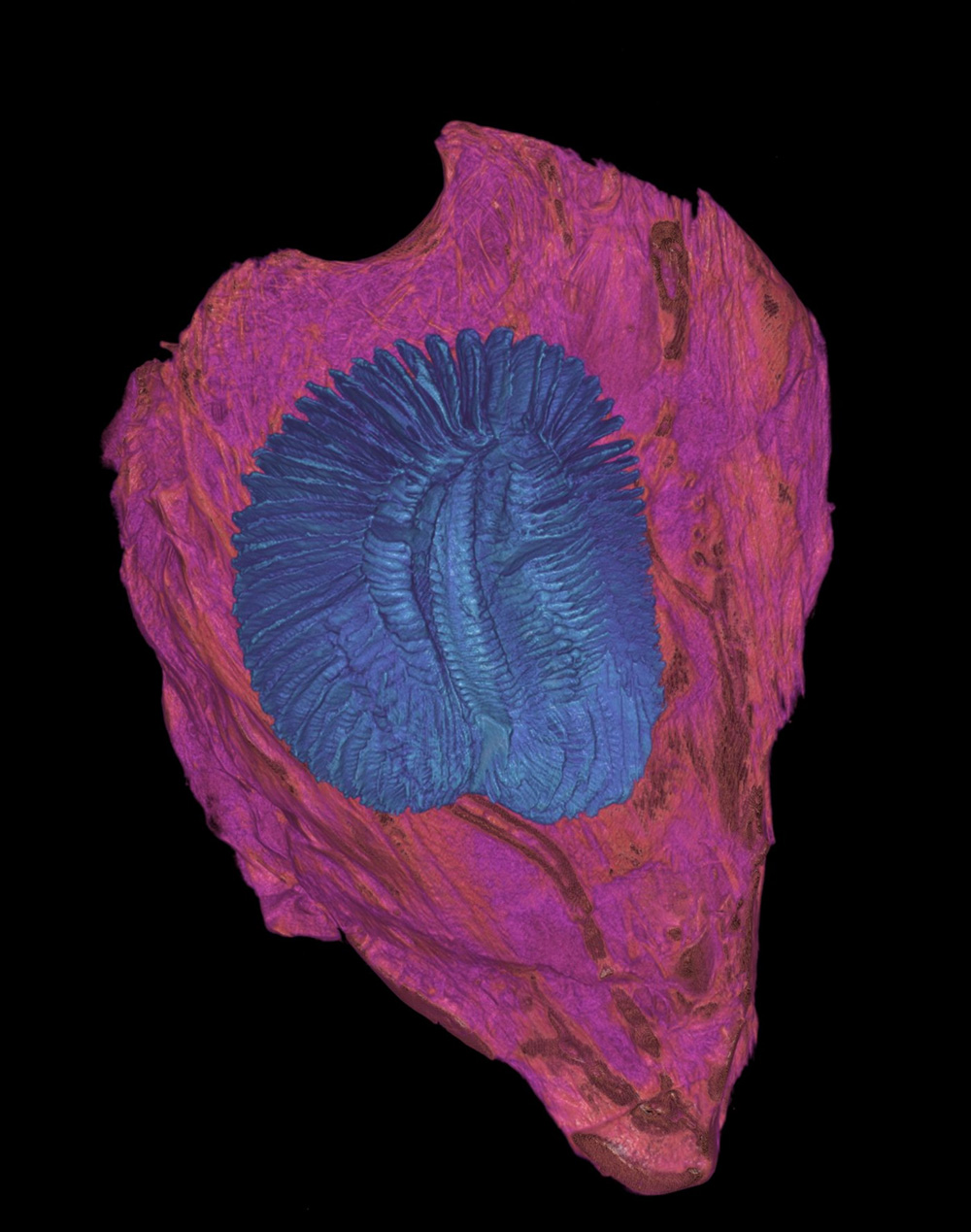
Honorable Mention: Hidden Current
FAU High School, Charles E. Schmidt College of Science
Mentor: Marianne Porter, Ph.D.
This image of a shortfin mako shark (Isurus oxyrinchus) olfactory rosette was imaged through contrast-enhanced micro-computed tomography (microCT). It shows a segmented rosette (blue) enclosed within its capsule (pink). The rosette, located within the snout, is responsible for odorant detection. It is composed of folded, plate-like tissue called lamellae, which increases the surface area for detecting odorants in the environment. Across different species, rosette morphology drastically varies, which could influence patterns of water flow and sensory function. The segmentation visible here highlights the structural organization of the rosette and lamellae in the shortfin mako. At the same time, the surrounding capsule provides protection and support. Image taken at the Berlin Family Bioimaging Lab at FAU Lab Schools Marcus Research and Innovation Center.