5/6/2026
Hidden Damage in Stony Corals
Study Uses 3D Imaging and AI to Find Hidden Damage in Stony Corals
Florida’s coral reefs are facing severe threats from diseases like Stony Coral Tissue Loss Disease (SCTLD), which has spread rapidly since 2014, killing large numbers of reef-building corals. This disease, along with others, weakens coral skeletons, reduces biodiversity, and threatens the overall health and resilience of reef ecosystems. Yet little is known about how these illnesses alter coral skeletons at the microscopic level. Understanding these structural changes is critical for monitoring reef health and guiding conservation efforts.
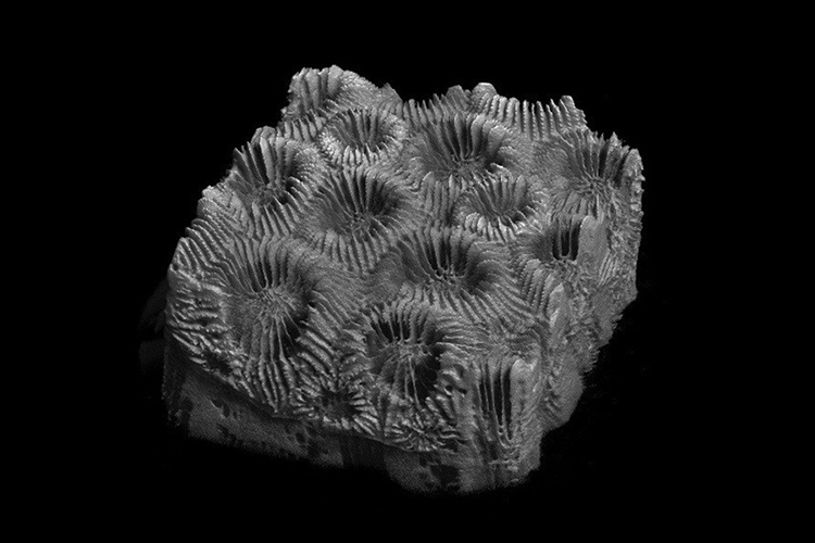
To address this challenge, FAU researchers used X-ray microcomputed tomography (micro-CT) combined with deep learning-based image segmentation to analyze coral skeletons in 3D. Focusing on healthy and SCTLD-affected specimens of hard stony corals, the team applied U-Net-based neural networks to automatically identify pores and skeletal structures. This approach allowed them to map porosity, density and thickness with 98% accuracy, providing a faster and more efficient way to assess how environmental stressors impact coral skeletons compared with traditional manual methods.
“Without high-resolution, 3D insights, scientists cannot fully understand how disease, warming oceans and other stressors compromise reef survival,” said Vivian Merk, Ph.D., corresponding author and assistant professor in the Department of Chemistry and Biochemistry in FAU’s Charles E. Schmidt College of Science and the Department of Ocean and Mechanical Engineering in the College of Engineering and Computer Science. “Our analyses provide a clearer, quantitative picture of how environmental stressors reshape coral skeletons at the microscopic level. By uncovering these hidden changes in porosity, density and skeletal thickness, we can see exactly how diseases like Stony Coral Tissue Loss Disease alter the physical integrity of corals.”