Instruments & Equipment
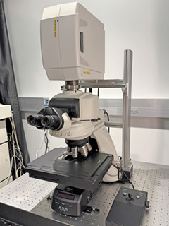
Fluorescent Microscopy
Location: FAU Jupiter
Overview:
Compact easy-to-use fluorescent microscope with high resolution monochrome
CCD
camera. With its fully motorized XYZ stage large samples can be captured and stitched fully automatically.
Confocal Microscopy
Location: FAU Jupiter
Overview:
The Nikon A1R confocal microscope is an easy to operate imaging system,
used for a
wide range of imaging applications. In addition to the four standard detectors, the system is equipped with
a spectral detector enabling acquisition of up to 32 channels simultaneously.
Location: FAU Jupiter
Overview:
The Nikon A1R confocal microscope system allows for the acquisition of
four
fluorescent channels simultaneously. For fast image acquisition the resonant scanner can achieve
scanning speeds of up to 420 fps. Mounted on an upright microscope stand, this is the preferred system
for fixed sample slides.
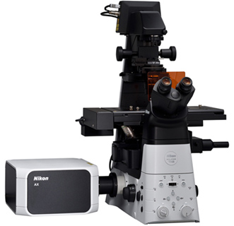
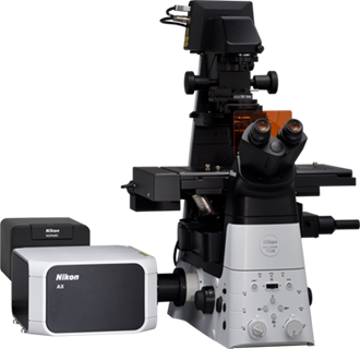
Location: FAU Jupiter
Overview:
The Nikon AXR confocal microscope combines fast scanning with a large
field of
view. Mounted on an inverted microscope stand and equipped with four laser lines and four detectors,
this system offers high versatility for a wide range of imaging applications.
Super-Resolution Microscopy
Location: FAU Jupiter
Overview:
The Nikon AXR with NSPARC imaging system provides high-resolution imaging
with
exceptionally low noise. Its specialized detector enables an improved signal-to-noise ratio, allowing
high-quality image acquisition at much lower excitation power. This helps preserve fluorescence and
enables gentler imaging of live samples.
Location: FAU Boca Raton
Overview:
This system combines confocal and super-resolution capabilities in one
setup.
Visualization of small intracellular structures and their function can be achieved using structured
illumination (SIM) technology. The N-SIM E realizes double the spatial resolution of conventional
optical microscopes (to approximately 115 nm)
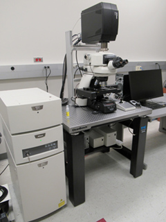
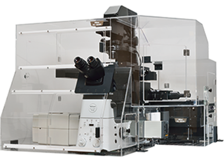
Multi-Photon Microscopy
Location: FAU Jupiter
Overview:
Nikon's A1R multiphoton microscope enables high speed, deep tissue imaging
in
live organisms with great sensitivity and clarity. The system features an ultra-high speed resonant
scanner capable of up to 420 fps.
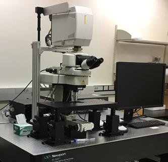
Light-Sheet Microscopy
Location: FAU Jupiter
Overview:
For fast acquisition of large samples such as whole mouse brains, organs,
or
other cleared tissues, the UltraMicroscope Blaze is equipped with LightSpeed Mode. This light-sheet
microscope is highly user-friendly and enables imaging of up to four different fluorescent channels.
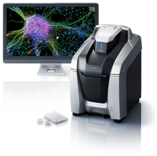
Digital Spatial Profiler
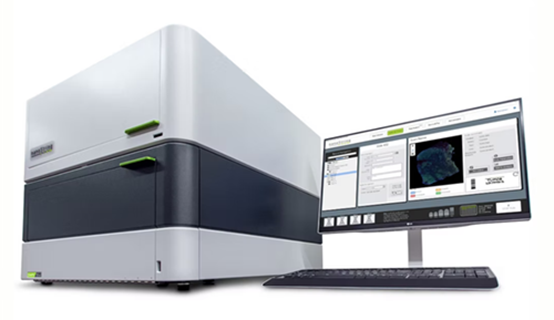
Location: FAU Jupiter
Overview:
The GeoMx DSP is a flexible and scalable platform for spatial biology.
GeoMX
is the only spatial biology platform that non-destructively profiles expression of RNA and protein
from distinct tissue compartments and cell populations with an automated and scalable workflow that
integrates with standard histology staining. Spatially profile the whole transcriptome and 570+
protein targets separately or simultaneously from your choice of sample inputs: whole tissue
sections, tissue microarrays (TMAs), or organoids.
Image Analysis Workstations
The Advanced Cell Imaging Core offers a variety of image analysis workstations for 2D and 3D image analysis. The following software tools are available free of charge for users of the Core:
Nikon NIS-Elements Advanced Research with 2D/3D Deconvolution module
Oxford Instruments Imaris 3D/4D Image Analysis Software
MBF Bioscience Neurolucida Neuron tracing and analysis software
MBF Bioscience NeuroInfo (whole brain investigation of circuits, neuronal populations, and biochemical marker expression)
Miltenyi Biotec MACS® iQ View – 3D Large Volume