
Great advancements in microscope technology enable leaps in neuroscience discovery – and they give us an inside view of the remarkable beauty of the brain.
Images of that beauty were recently recognized in Florida Atlantic University’s Art of Science photography and videography competition. The annual contest was created to celebrate the intersection of creativity and visual beauty of scientific research, inviting faculty, students and staff to showcase their work. The competition highlights how science and art collide, strengthening connections between the research community and the public while reflecting FAU’s growing reputation for innovation, discovery and excellence.
This year’s competition drew a record-breaking 200 entries across disciplines including neuroscience. On the next few pages, enjoy a close-up look at some of the winners from the Stiles-Nicholson Brain Institute.
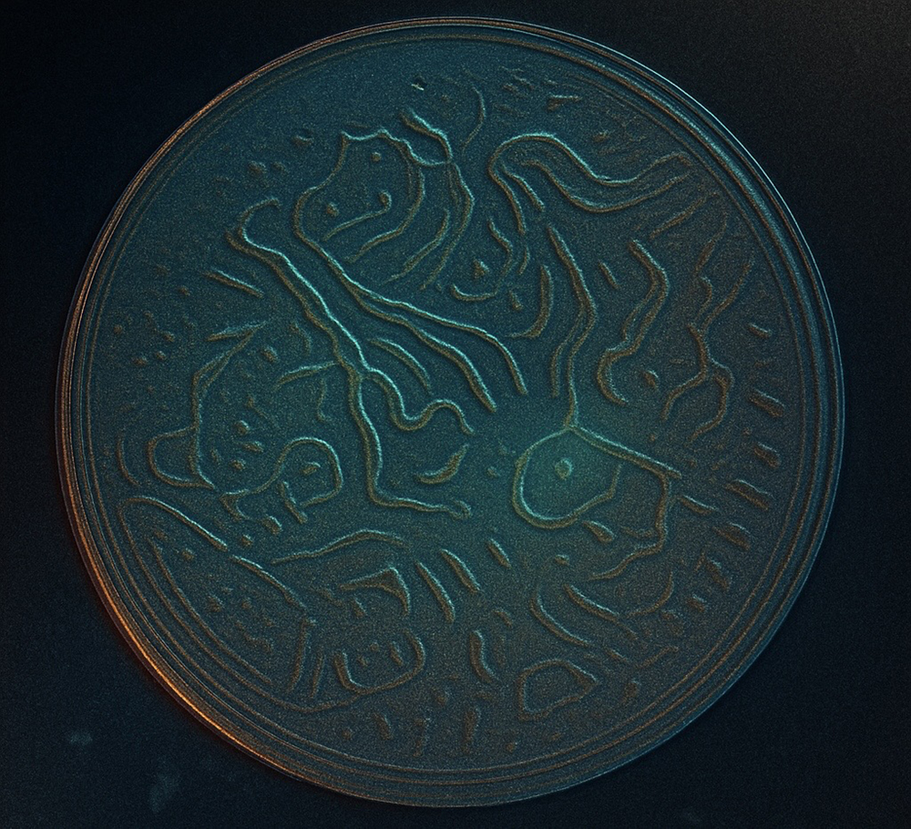
TOP POSTDOC
Hussam Alshareef, Ph.D., postdoctoral fellow, Stiles-Nicholson
Brain Institute
Here, the C. elegans nematodes were left searching for more food, and instead of hiding their need, they etched their hunger into the agar, leaving behind an abstract pattern. What looks like art is actually survival - a biological request written in unexpected lines.
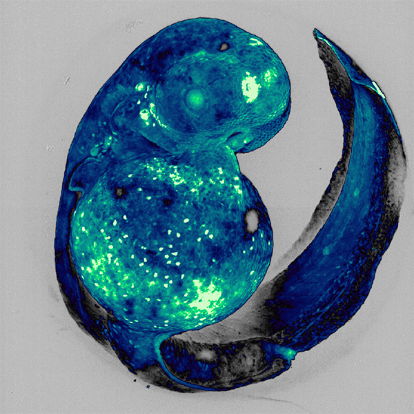
RESEARCH EXCELLENCE
Tessa Dallo, doctoral student, Charles E. Schmidt College
of Science and Stiles-Nicholson Brain Institute Mentor: Laura Fontenas, Ph.D.
This image captures a developing zebrafish embryo, a powerful model for exploring how the nervous system takes shape. The embryo is curled as it has not yet hatched. It expresses zebrabow, a genetic tool that fluorescently labels many cells, enabling researchers to visualize cell development and interactions in vivid detail. Because zebrafish embryos are transparent, they provide a window into the earliest stages of life, when the foundations of movement and sensation first emerge. The image has been falsely colored for artistic effect, highlighting the intricate beauty of developing structures that guide our understanding of both healthy and disordered development.
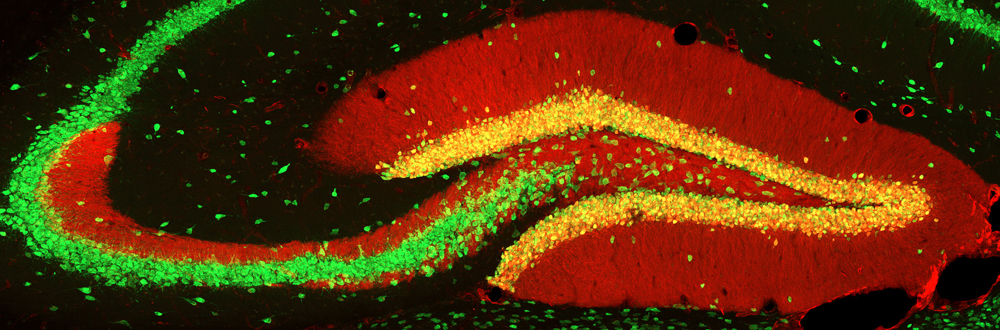
HONORABLE MENTION
Marianne Charlene Monet, graduate student, Charles E. Schmidt
College of Science and Stiles-Nicholson Brain Institute
Mentor: Ning Quan, Ph.D.
This image shows part of the brain where memories begin their journey, the hippocampus, a region essential for learning and memory. The red glow highlights a special group of neurons known as granule cells, located in an area called the dentate gyrus. What makes this image unique is the long red “tail” stretching from these cells into other hippocampus regions called CA2 and CA3. This vibrant projection reflects how the brain encodes, organizes and communicates new memories. The green signal, labeled brain cells with NeuN, marks other neurons including pyramidal cells in CA2/3, which are involved in processing and retrieving social memories. Within these neural circuits, the brain begins to transform experience into memory and forms the foundation for how we recall events, recognize others and navigate the social world around us.