State-of-the-Art Microscope Blazes New Trail in Brain Imaging
by Bethany Alex | Thursday, Aug 26, 2021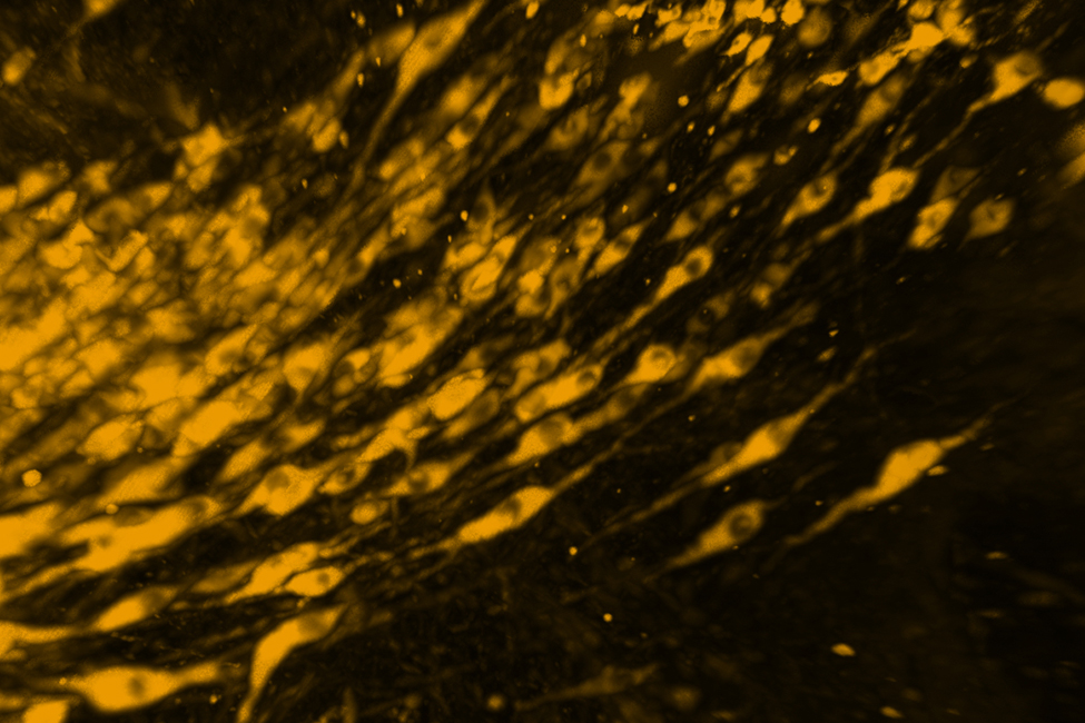
Scientists at FAU's Stiles-Nicholson Brain Institute are among the first in the state of Florida to capture 3D images of clusters of identified brain cells in the intact brain using a new, fully automated, microscope capable of imaging preparations with a razor-thin sheet of laser light. The microscope – also known as the "Miltenyi UltraMicroscope Blaze" – is a strategic investment by the Institute to provide advanced imaging opportunities for FAU neuroscientists and their trainees.
"The Blaze is the first automated light sheet microscope in South Florida, and one of only two in the state," said Randy D. Blakely, Ph.D., executive director of FAU’s Stiles-Nicholson Brain Institute. "It's really going to accelerate our science and add to the tremendous technical resources FAU offers its scientific community. We have been eyeing this technology for some time, seeing the microscope’s capabilities as transformative, allowing us to look deep into the brain with molecular precision and circuit level resolution without needing to disrupt the normal structure of the tissue."
In short, the new microscope’s large field of illumination – a sheet of light rather than a point of light typical of other microscopes – allows for intact brains of mice to be imaged down to the subcellular level at high speed and with neuronal connections intact. Such visualizations are essential in understanding how diseases impact brain architecture and how global activity patterns are formed.
"The Blaze has the ability to provide us with a bird's-eye view of whole organs without losing subcellular details, something biologists have been longing for," said Qi Zhang, Ph.D., Cell Imaging Core Scientific director and research associate professor for FAU's College of Medicine. Dr. Jana Strickler, managing director of the Core, is now training scientists to use the new instrument and analyze the large datasets it can create. In 2016, the Institute’s commitment to high-end imaging technologies landed FAU a prestigious Nikon Center of Excellence.
“The Jupiter campus is home to terrific research facilities, not only at FAU, but also at the Max Planck Florida Institute for Neuroscience, well-known for its electron microscopy and super- resolution imaging resources, and at the nearby Florida home of Scripps Research”, said Blakely. “With the Blaze, we take a big step forward in understanding the amazing complexity of the brain.” Watch a video of dopamine and norepinephrine neurons in the mouse brain courtesy of Dr. Lorena Areal, Dr. Paula Kurdziel and Dr. Jana Strickler, Managing Director of the Cell Imaging Core.