Study Uncovers Mechanics of ‘Tail-Whipping’ in Thresher Sharks
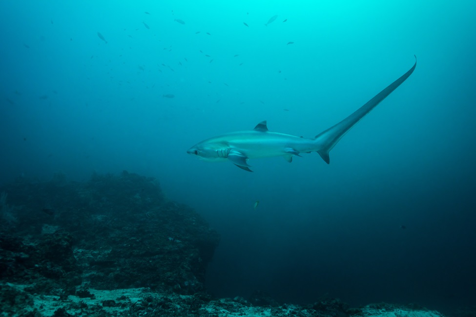
The tresher shark's exceptionally elongated tail can often be as long as their entire body,
Like Indiana Jones, thresher sharks (Alopias spp.) have mastered the art of the whip using their tails. With incredible speed, their long, machete-like tails can slap and stun their prey, allowing them to swallow multiple fish in one fell swoop. Their exceptionally elongated tail, which can often be as long as their entire body, not only makes this particular shark unique, but also a formidable hunter.
Thresher shark “tail-whipping” consists of four phases: preparation, strike, wind-down recovery, and prey collection. Overhead tail slaps begin in the preparation phase by lunging toward targeted prey. The strike phase begins by lowering the head and flexing the body, which raises the tail over their head to create a whip-like motion. The wind-down recovery phase consists of the shark returning to swimming posture, and consuming stunned prey.
The shark’s tail-whipping movement dramatically differs from the side-to-side motion produced by its body during swimming. The cartilaginous vertebral column, which is the main body axis, may have anatomical modifications to withstand the extreme bending during the tail-whipping behavior. Prior research has examined the vertebrae of thresher sharks, but in the context of the forces experienced during swimming.
Now, new research from Florida Atlantic University in collaboration with Apex Predators Program, Northeast Fisheries Science Center, National Oceanic and Atmospheric Administration (NOAA), provides intricate details showing that anatomy of the vertebrae might support the mechanics of extreme body bending in thresher sharks, enabling these expert hunters to weaponize their tails.
For this study, researchers examined vertebrae from the head and tail ends of the body. They investigated vertebral anatomy and measured variables like height, width and length along the vertebral column from 10 common thresher sharks (Alopias vulpinus) across a range of sizes from an embryo to large adults.
Researchers used micro-CT scanning, similar to CAT scans humans use in medical facilities, to image the internal architecture of each vertebra and quantified various mineral structures found in the calcified cartilage. They also used two-dimensional shape analysis techniques to examine variation in the spatial distribution of mineral structures along the vertebral column.
Results of the study, published in the journal Royal Society Open Science , suggest thresher shark vertebral anatomy and mineralized microstructure meet the demands required for fast swimming and tail-whipping behavior seen in these species.
Researchers found that the mineralized microarchitecture in thresher shark vertebrae changes in the front and the back of the body, and these anatomical modifications may support their unique tail-whipping behavior. Essentially, thresher shark vertebral column is fortified along its length and might work like a catapult, allowing the tail to launch over the head.
“We found that anatomy and microstructure significantly varied along the body and among developmental groups – embryonic, juvenile and adult common thresher sharks,” said Jamie L. Knaub, first author and a doctoral graduate student in FAU’s Department of Biological Sciences within the Charles E. Schmidt College of Science. “Based on our results, we believe that thresher shark vertebrae vary in anatomy, and the amount and arrangement of mineral, supporting the mechanical needs for tail-whipping.”
The researchers also discovered that juvenile-sized sharks acquire mineralized structures throughout development, likely to support a larger body as they grow and tail for whipping behaviors.
“We think that anterior body vertebrae stabilize the thresher shark’s main body, while vertebrae closer to the tail support overhead tail-whips,” said Marianne E. Porter, Ph.D., senior author and an associate professor, FAU Department of Biological Sciences. “Additionally, developmental changes suggest that vertebral anatomy shifts across development to support a larger body and caudal fin.”
Thresher sharks, part of the Alopiidae family, comprise three species: the pelagic thresher (Alopias pelagicus), the bigeye thresher (Alopias superciliousus), and the common thresher (Alopias vulpinus). All three thresher shark species have been listed as vulnerable to extinction by the World Conservation Union since 2007.
Study co-authors are Michelle Passerotti, Ph.D., a fish biologist, Apex Predators Program, NOAA Fisheries; Lisa J. Natanson, Ph.D., a shark researcher, Apex Predators Program, NOAA Fisheries (retired); and Tricia Meredith, Ph.D., director of research, FAU A.D. Henderson University School and FAU High School, and an assistant research professor, FAU College of Education.
The work was conducted as part of Knaub’s Ph.D. research, which was supported by a National Science Foundation (NSF) CAREER grant awarded to Porter (NSF, IOS-1941713), the Jim Elliot Award from Tomography for Scientific Advancement awarded to Knaub, and the Vincent Saurino Fellowship, the Newell Doctoral Fellowship, and the National Save the Sea Turtle Foundation Scholarship from FAU awarded to Knaub.
-FAU-
Tags: technology | faculty and staff | students | science | research