FAU Workshops
Workshop on Light Microscopy & Cellular Imaging in Life Sciences
Course Description
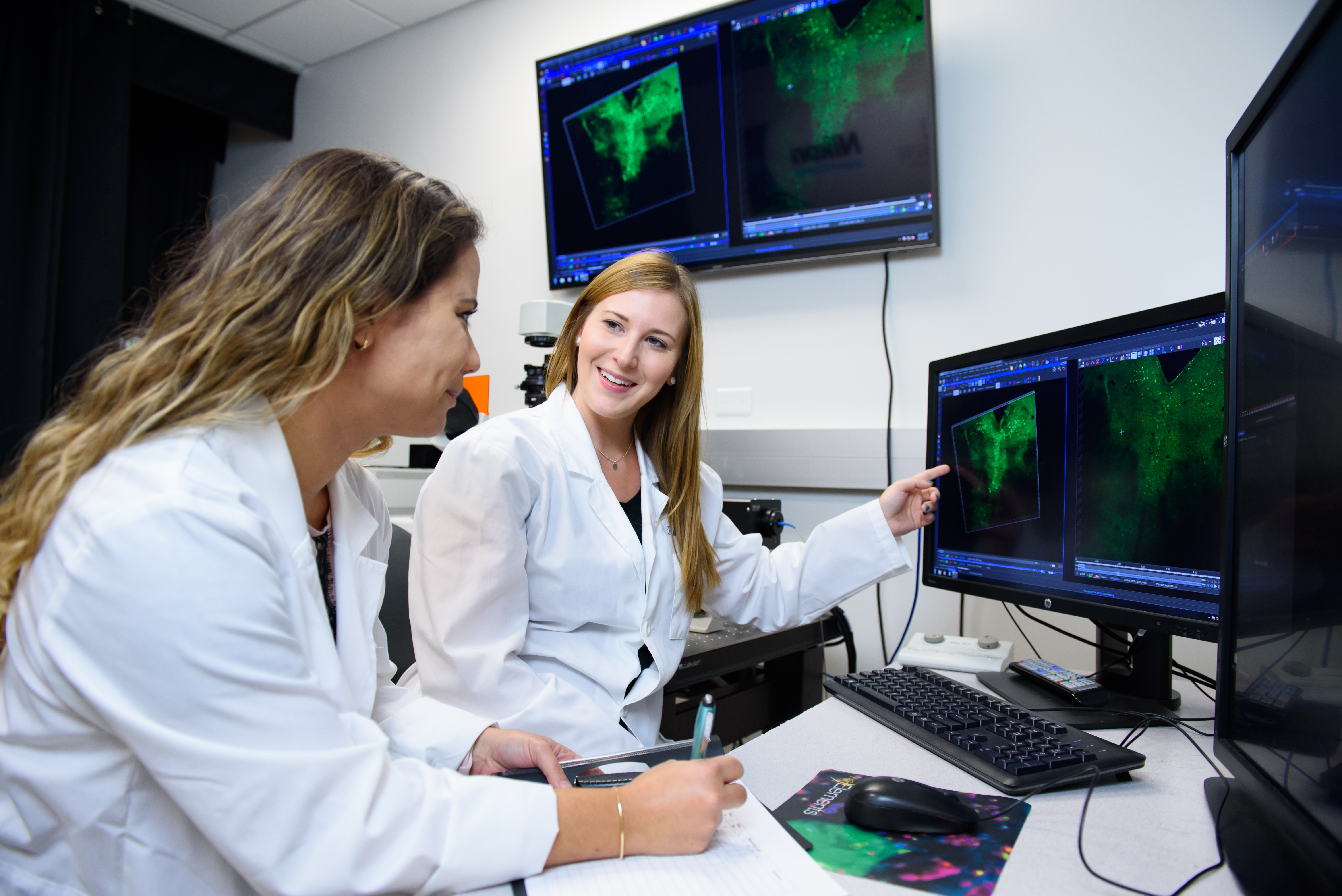
An intensive, three-day workshop in light microscopy for trainees in the life sciences. The course helps students understand the theory of image formation and give an introduction to different fluorescent and laser scanning techniques. Practical sessions include demonstration of imaging systems available in the FAU Cell Imaging Core.
The course consists of lectures in the morning and practical demonstrations in the afternoon. During the pratical sessions, different microscope techniques are demonstrated using sample preparations.
Lectures
Image formation (lenses and image formation, objectives, diffraction, point spread function, resolution)
- Fluorescence microscopy (fluorescent probes and proteins, laser, confocal microscopy, two-photon microscopy, super-resolution microscopes)
- Detection and image analysis (cameras and detectors, digital images, image analysis, deconvolution)
Practical Sessions and Demonstrations
- DIC/Wide-field/Fluorescent microscopy Confocal microscope, image acquisition of fixed samples (Z-stacks, multi-channel)
-Confocal microscope, live cell imaging (time-lapse, FRAP, calcium imaging)
-Multi-Photon microscope (laser ablation) Image and data analysis in NIS Elements, ImageJ, Amira, and Neurolucida
-Students are encouraged to bring their own samples for imaging
Special Demonstrations
Representatives from NanoLive, Keyence, and Mizar Imaging will join us for special demonstrations of their newest technology.
- NanoLive: 3D Cell Explorer-fluo
- Keyence: BZ-X800
- Mizar Imaging: Tilt - High-Resolution Light Sheet Imaging
Contact: Jana Boerner, FAU Cell Imaging Core, Managing Director - jboerner@fau.edu