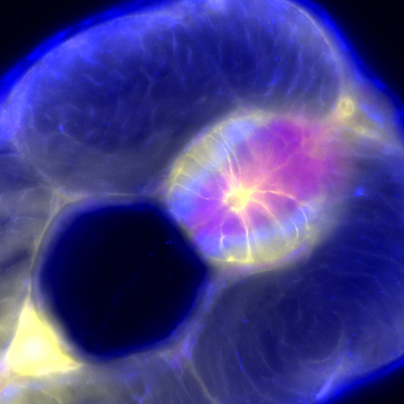
First Place: Zebrafish Spinal Cord
Photography by Addison Manofsky, undergraduate student,
Harriet L. Wilkes Honors College and FAU High School
This is an image of a transverse section of a zebrafish embryo. The large, dark circle is the notochord, which will be replaced by the vertebral column. The bright area beside it, filled with several branches of neurons and various glia, is the spinal cord. In both the peripheral and central nervous system, glial cells play an important role in supporting and myelinating neurons. Muscles, in addition to glia, can be seen in the periphery surrounding the notochord and spinal cord. Imaging aids in the study of glial migration, development and myelination, allowing for the research of glial cells and their relations to neurological conditions.
Mentor: Laura Fontenas, Ph.D., assistant professor, Charles E. Schmidt College of Science