Florida Atlantic Society for Neuroscience Meeting 2025
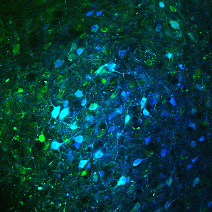
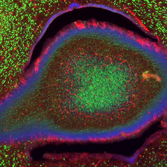
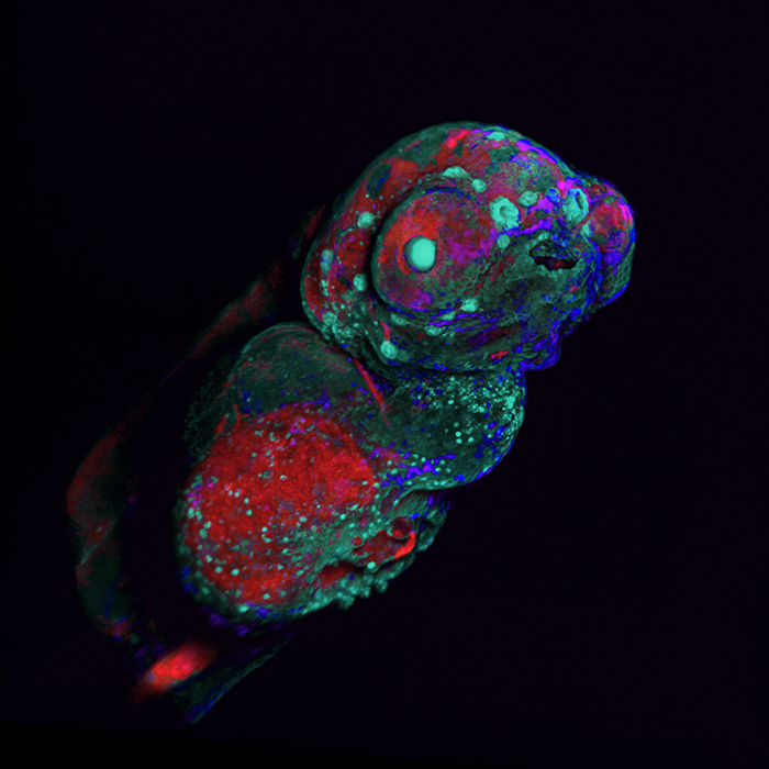
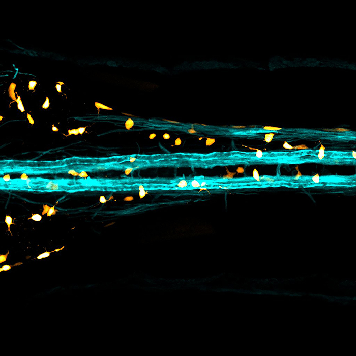
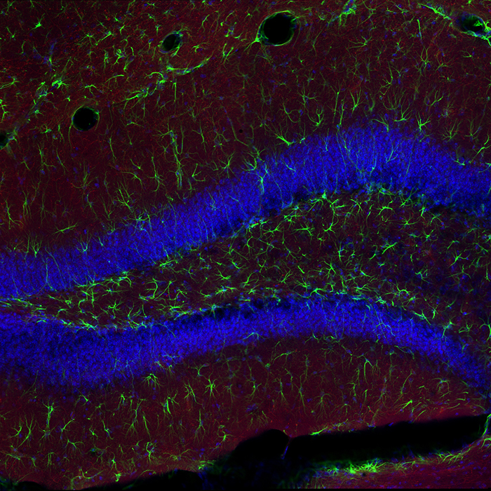
Viewfinder images
by Marianne Charlene Monet, graduate student, Charles E. Schmidt College of Science
“In my research, I study the distribution of IL-1R1, a receptor in the developing brain. While working with a 14-day-old mouse, I captured this stunning image from the cerebellum, a part of the brain I don't usually focus on. What's fascinating is that it looks like an eye! In the image, the red color highlights blood vessels and neurons that have IL-1R1, while the green marks all the neurons. Although I don't study this region in detail, the beauty of this image really caught my attention, and I felt compelled to share it with others.”
by Tessa Dallo, doctoral student, Stiles-Nicholson Brain Institute
Using ubiquitously expressed Zebrabow, a genetic tool aptly named for its diverse color profile, we can use confocal imaging to capture a wide array of fluorescently labelled cells. By injecting specific RNA into zebrafish embryos at the one-cell stage, a recombinase facilitates combinatorial expression of red, yellow, and cyan florescent proteins resulting in a range of colorful hues. After only a few days, we can see the rainbow overlay of different cells. Since cells with the same origin will have the same level of recombination, and therefore the same color, this is a valuable tool that can allow us to conduct cell lineage analyses.
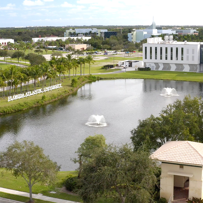
Florida Atlantic University’s John D. Macarthur Campus in Jupiter is where groundbreaking programs in research and education create unmatched opportunities for the best and brightest students. It’s where FAU converges on site with two of the world’s leading research organizations, the Max Planck Florida Institute for Neuroscience and The Herbert Wertheim UF Scripps Institute for Biomedical Innovation & Technology, to offer high school, undergraduate and graduate students transformational experiences not found anywhere else in the world.
by Laura Fontenas, Ph.D., assistant professor, Charles E. Schmidt College of Science
Glia are essential cells that partner with neurons to ensure the proper function of the nervous system. This picture represents pseudo-colored glia in and out of the spinal cord of a zebrafish embryo.
by Paula Kurdziel, Ph.D., research assistant professor, Stiles-Nicholson Brain Institute and Harriet L. Wilkes Honors College
Hippocampal granule cells are represented in blue, glial cells represented in green and red coloring shows serotonergic fibers by labeling the serotonin transporter.
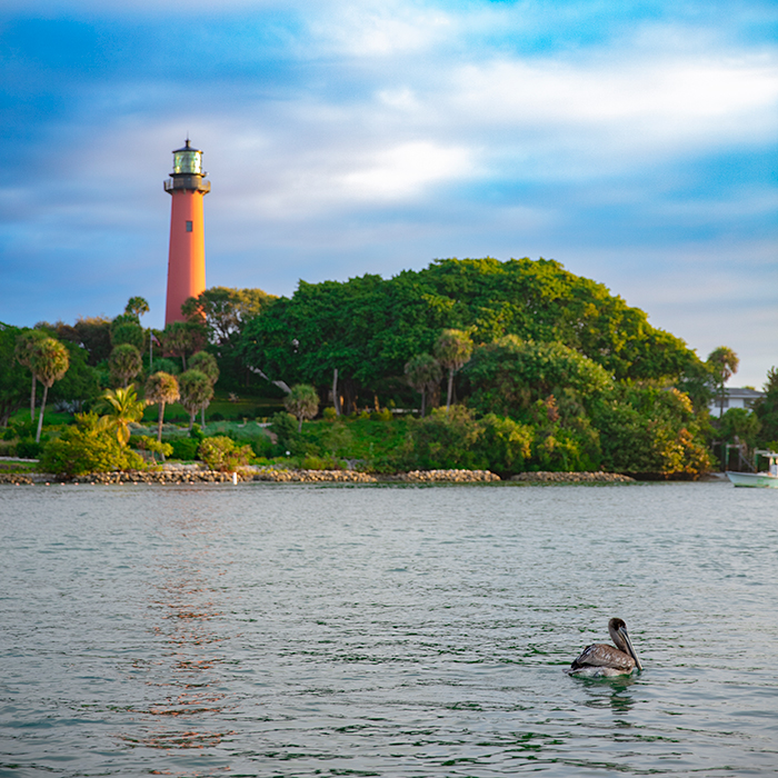
The Jupiter Inlet Lighthouse located in Jupiter, Fla., on the north side of the Jupiter Inlet. The site for the lighthouse was chosen in 1853 and was constructed in 1860 and has had more than 70 lighthouse keepers that served from 1860 to 1939.
by Tim Holford, Ph.D., Florida Atlantic Max Plank Honors Program, Charles E. Schmidt College of Science, Max Planck Florida Institute for Neuroscience and Harriet L. Wilkes Honors College
Neurons in the central amygdala are important for regulating various aspects of social and emotional life and are commonly disrupted in neurological disorders. In our research, we identify circuit abnormalities in a mouse model of autism spectrum disorder where the regulatory protein PTEN is removed from a subset of inhibitory cells in the brain called Somatostatin-expressing interneurons. Mice carrying the SOM-PTEN deletion displayed elevated levels of fear expression, anxiety and sensory sensitivity, as well as substantially impaired connectivity and synaptic strength within the central amygdala compared to wild type mice, which could lead to those behavioral disruptions. This image was acquired during a circuit mapping experiment, where we probe cell to cell communication using a combination of 2-photon microscopy, optogenetics and electrophysiology to stimulate and image individual neurons while recording their influence onto a postsynaptic cell. SOM cells are blue and GCamp cells (to read out neural activity) are green.