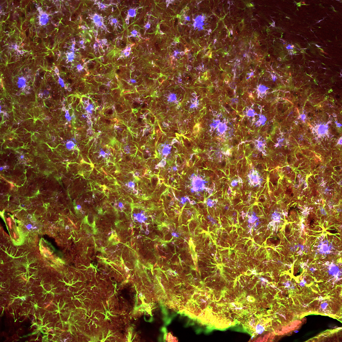
Aged 5xFAD Alzheimer's Mouse Hippocampus - Hippocampal brain sections of a transgenic Alzheimer's mouse model of a 6-month-old mouse were investigated through IHC antibody staining such that the final antibody added will reflect a colored wavelength: Amyloid Beta Plaques (blue: Amylo Glo), Astrocytes (green: GFAP), IL1R1 (red: tdTom), Microglia (far red/gray: IBA1). At this stage in the mouse's lifespan Alzheimer's Disease pathology has progressed to the point of causing behavioral changes and cognitive deficits.
Microglia and Astrocytes, the amyloid beta response team, are recruited and activated to engulf and dispose of the amyloid beta plaques. Both of these cell types begin to wrap and surround the plaques with their cell processes as amyloid beta is accumulated during Alzheimer's pathology progression. Images were captured on a confocal microscope by creating a z-stack image (multiple layers pressed together) and edited on Biorender.