Octopus Americanus
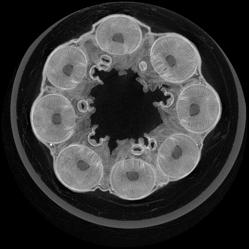
Micro-CT images taken of Octopus Americanus for ongoing research on comparative morphology between local octopus species. Micro-CT offers a non-invasive method of visualizing internal morphology in 3D. This is a cross section directly below the head of the octopus. Each of the eight arms are visible, along with the axial nerve cord in the middle of each arm. Images were taken at the Berlin Family Bioimaging Lab at FAU Lab Schools Marcus Research and Innovation Center.