Fruit Fly Desire
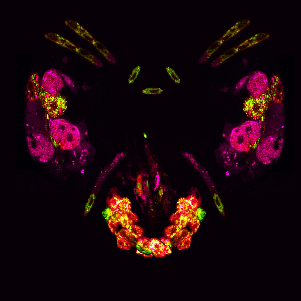
The image depicts the Insulin Producing Cells (IPCs) and Diuretic Hormone 44 (DH44) neurons of
Drosophila melanogaster (the common fruit fly), arranged to resemble the fly's head. Initial images were
captured using a 40x objective lens on a confocal microscope. Most of the head structure was comprised of a
13-day old, starved female Drosophila melanogaster, while the lower proboscis was an eight- to nine-day-old
male fly that was fed on a glucose and sucrose rich diet. The cells were genetically modified to express two
different fluorescent markers: Drosophila Distracted protein in pink and Drosophila insulin like peptides in
green. IPCs (pink) are analogous to mammalian pancreatic beta cells, which release insulin or insulin-like
hormones to regulate high blood sugar levels. DH44 neurons (green) are analogous to mammalian
corticotrophin-releasing hormone (CRH) which play a crucial role in detecting dietary amino acids (the
building blocks of proteins) and promote food searching and consumption behaviors. Both IPCs and DH44
neurons play essential roles in maintaining metabolic homeostasis — keeping the body's internal environment
balanced to support optimal metabolic function. Dysfunction in these cell types can contribute to metabolic
disorders such as diabetes and obesity, as well as increase the risk of neurodegenerative diseases.
This project aims to uncover the mechanisms that drive endolysosomal trafficking — the process which vesicles,
called endosomes, transport cellular material to lysosomes for sorting, recycling, or disposal — with a focus
on the Distracted protein