Coral Coded
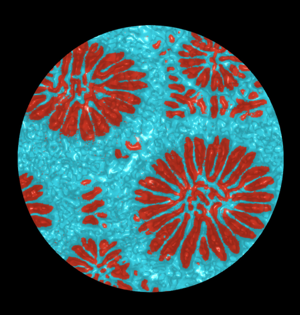
Coral skeletons may appear as solid rock, but under the surface they have porous networks that play a role in their growth and survival strategies. To study this hidden architecture, we use X-ray micro-computed tomography (micro-CT), similar to medical CT scans, to analyze the 3D volumes of these coral skeletons. We are then able to efficiently separate the solid skeleton and pores of these large datasets using Artificial Intelligence (AI). This process, called segmentation, gives us a clear map of both the solid structure and the porosity within. By turning these complex images into colorful, easy-to-read data, we can better understand how coral skeletons are built, how strong they are, and how they may respond to environmental stress and disease. This image is a 2D slice of a Montastraea cavernosa coral skeleton, illustrating the porous radial septal regions in orange, surrounded by skeleton in the thecal regions in blue. Using deep-learning AI models, an image stack of 2000 images from a 3D scan can be segmented in a matter of a few hours with high accuracy, much faster than a conventional, manual segmentation. Micro-CT scans were collected at the Berlin Family Bioimaging Lab at FAU Lab Schools Marcus Research and Innovation Center.