Equipment
Located in In the College of Medicine on Boca campus, Nikon’s N-SIM E system with A1+ confocal unit, combines confocal and super-resolution capabilities in one system. Visualization of small intracellular structures and their function can be achieved using structured illumination (SIM) technology. N-SIM almost doubles the resolution of conventional confocal microscopy.
On Jupiter campus, within the research facilities of the Jupiter Life Science Initiative and the Brain Institute, the Nikon A1R MP multiphoton microscope, is an excellent system for in vivo high-speed scanning of signals deep within the living animal and intact brain tissue. Further, the institutions house two Nikon confocal systems. The Nikon C1 confocal system with spectral detector, is a basic confocal imaging system. The Nikon A1R confocal system is equipped with six laser lines, spectral detector, and a TIRF imaging system. Nikon’s laser TIRF system yields an extremely high signal to noise ratio and allows for observing single molecules in living cells. The Keyence All-in-One Fluorescence Microscope, is an easy-to-use, automated system for imaging a variety of samples, from live cells to fixed brain sections.
Fluorescent Microscopy
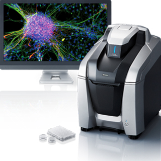
Location: FAU Jupiter
Overview:
Compact easy-to-use fluorescent microscope with high resolution monochrome CCD camera. With its fully motorized XYZ stage large samples can be captured and stitched fully automatically.
- Inverted fluorescence phase contrast microscope
- Bright field, Fluorescence, and Phase contrast observation modules
- Electronic XYZ stage
- Fluorescent filters for DAPI, GFP, and TexasRed
- 2.83 million pixel monochrome CCD camera, 15 fps for monochrome recording, 4,080 x 3,060 max pixel resolution
- Multi-color image capturing capability
- Sectioning software module for Z-stack image capturing
- Objectives:
- Plan Apo λ 2x
- Plan Apo λ 10x
- Plan Fluor 20x
- S Plan Fluor ELWD 40x
- Plan Apo λ 60x Oil
Confocal Microscopy
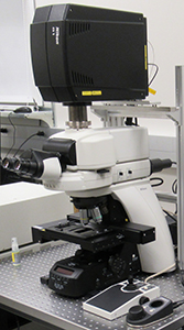
Location: FAU Jupiter
Overview:
The Nikon A1R confocal microscope is an advanced confocal imaging system used for a wide range of imaging applications. Mounted on an upright microscope with motorized stage and equipped with Nikon’s newest resonant scanner it makes imaging of large samples quick and easy.
- Four laser lines: 408, 488, 561, 640 nm
- Four detectors: blue, green, red, far red; with two high sensitivity GaAsP detectorsv
- High-resolution Galvano scanner with pixel size up to 4096 x 4096; scan speed 2 fps at standard mode and 10 fps in fast mode (at 512 x 512 pixels, bi-direction)
- High definition 1K resonant scanner with maximum resolution of 1024 x 1024 pixels (15 fps) and scanning speed up to 420 fps (at 512 x 32 pixels)
- Motorized XY stage
- Objectives:
- Plan Apo 10x
- Plan Apo 20x
- Plan Fluor 40x Oil
- Plan Apo 60x Oil
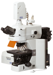
- Location: FAU Jupiter
Overview:
The A1R confocal microscope is a high-quality imaging system with a high-resolution Galvano scanner and high-sensitive GaAsP detectors. With its fast resonant scanner (up to 420 frames per second) high-speed 3D scanning can be achieved for live imaging. The A1R is additionally equipped with Nikon’s laser TIRF system, allowing for single molecule visualization and dynamic particle tracking.
- Fully motorized XY stage
- Six laser lines: 408, 455, 488, 514, 561, 640
- Four detectors: blue, green, red, far red; with two high sensitivity GaAsP detectors
- Spectral detector (maximum 32 channels, 400-700 nm range)
- High-resolution Galvano scanner with pixel size up to 4096 x 4096; scan speed 2 fps at standard mode and 10 fps in fast mode (at 512 x 512 pixels, bi-direction)
- High definition 1K resonant scanner with maximum resolution of 1024 x 1024 pixels (15 fps) and scanning speed up to 420 fps (at 512 x 32 pixels)
- Hamamatsu ORCA-Flash4.0 camera
- Laser TiRF Imaging system
- Motorized Piezo Z stage for high-speed Z-direction scanning
- Plan Apo λ 2x
- Plan Apo λ 4x
- Plan Apo λ 10x
- Plan Apo VC 20x DIC N2
- APO LWD 20x WI λ S
- Plan Apo TIRF 60x Oil DIC H N2
- Plan Apo λ 60x Oil
- Tokai Hit Stage top Incubator with temperature and CO2 control
- Objectives:
Super-Resolution Microscopy
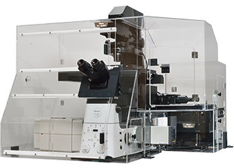
Location: FAU Boca Raton (College of Medicine)
Overview:
This system combines confocal and super-resolution capabilities in one setup. Visualization of small intracellular structures and their function can be achieved using structured illumination (SIM) technology. The N-SIM E realizes double the spatial resolution of conventional optical microscopes (to approximately 115 nm).
- Fully motorized XY stage
- Motorized Piezo Z stage for high-speed Z-direction scanning
- Four laser lines (408, 488, 561, 640)
- Four detectors: blue, green, red, far red; with two high sensitivity GaAsP detectors
- High-resolution Galvano scanner with pixel size up to 4096 x 4096; scan speed 2 fps at standard mode and 10 fps in fast mode (at 512 x 512 pixels, bi-direction)
- High definition 1K resonant scanner with maximum resolution of 1024 x 1024 pixels (15 fps) and scanning speed up to 420 fps (at 512 x 32 pixels)
- Hamamatsu ORCA-Flash4.0 camera
- Objectives: Plan Apo λ 10x
Plan Apo VC 20x
Plan Fluor 40x Oil
Apo 60x λS Oil
SR HP Apo TIRF 100x Oil
- Tokai Hit Stage top Incubator with temperature and CO2 control
- N-SIM analysis software
Multi-Photon Microscopy
Location: FAU Jupiter
Overview:
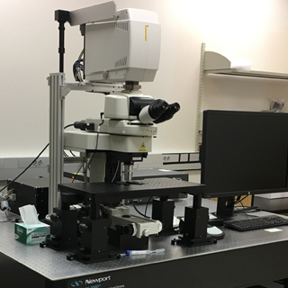
Nikon's A1R multiphoton microscope enables high speed, deep tissue imaging in live organisms with great sensitivity and clarity. The system features an ultra-high speed resonant scanner capable of up to 420 fps.
- Upright FN1 microscope
- Prior high precision motorized ZDeck Quick Adjust Platform System
- Coherent Chameleon Vision II Ti:Sapphire tunable laser (680 to 1080 nm)
- 450 µm Piezo-Z nosepiece for high-speed Z-stack acquisition
- Hybrid scanner head with ultrahigh-speed resonant scanner and high-resolution Galvano scanner
- Four-channel high sensitivity GaAsP detector
- 25x CFI APO LWD, 1.1 NA, 2 mm WD Water Immersion Objective
Image Analysis Workstations
Location: FAU Jupiter
Overview:
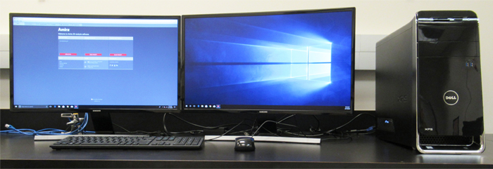
- Dell Desktop with NIS-Elements Advanced Research 5.0 and Deconvolution module
- Dell Desktop with Amira 3D Software
- Dell Desktop with Amira 3D Software and NIS-Elements Advanced Research 4.5
- Linux Desktop with NIS-Elements Advanced Research 5.0 and FRET analysis module;
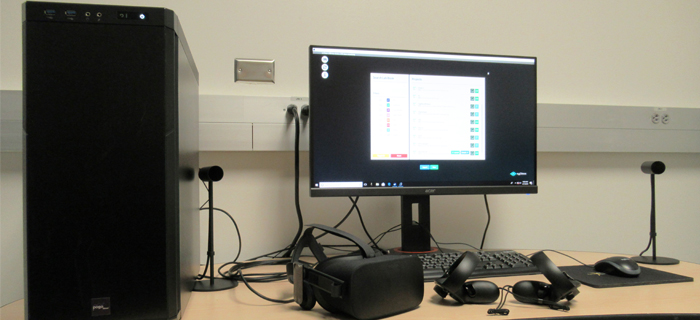
- SyGlass Visualization System with Oculus rift VR system
- HP Desktop with NIS-Elements Advanced Research 5.0 and SIM module